


Illustrated by Elena Nikonova
Surprisingly rapid regrowth of unused brain connections after decades of near blindness
Since 2007, clinical trials using gene therapy have resulted in often-dramatic sight restoration for dozens of children and adults who were otherwise doomed to blindness. Now, researchers from the Perelman School of Medicine at the University of Pennsylvania and The Children’s Hospital of Philadelphia (CHOP), have found evidence that this sight restoration leads to strengthening of visual pathways in the brain, published this week in Science Translational Medicine.
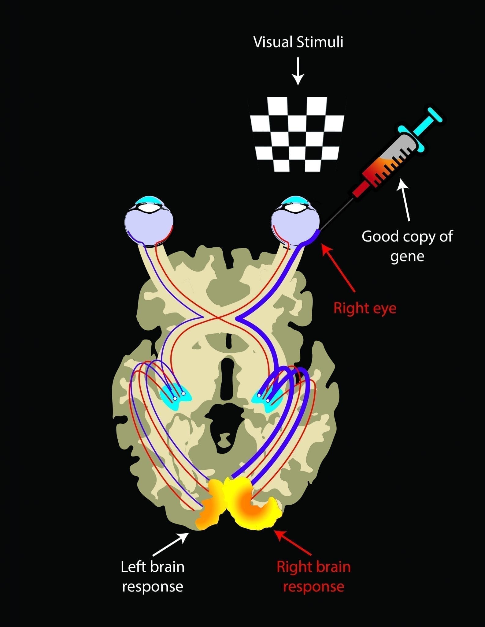
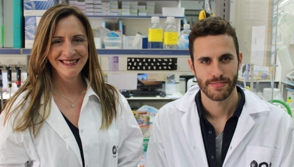
“The patients had received the gene therapy in just one eye (their worse seeing eye), and though we imaged their brains only about two years later, on average, we saw big differences between the side of the brain connected to the treated region of the injected eye and the side connected to the untreated eye,” said lead author Manzar Ashtari, PhD, director of CNS Imaging at the Center for Advanced Retinal and Ocular Therapeutics in the Department of Ophthalmology at Penn. Ashtari is the former director of Diffusion Tensor Image Analyses and Brain Morphometry at CHOP.
“It’s an elegant demonstration that these visual processing pathways can be restored even long after the period when it was thought there would be a loss of plasticity,” said senior author Jean Bennett, MD, PhD, the F.M. Kirby Professor of Ophthalmology at Penn and director of the Center for Advanced Retinal and Ocular Therapeutics.
The team examined ten patients who have Leber’s congenital amaurosis Type 2 (LCA2), a rare disease that afflicts those who inherit a bad copy of an LCA2 gene from each parent, LCA2 causes their retinas to degenerate slowly and they typically have limited visual function at birth and then experience progressive loss of their remaining vision, rendering them completely blind by mid-life.
In one of the first great success stories for gene therapy, Bennett’s team and others demonstrated the effectiveness of LCA2 gene augmentation in animal tests stretching back to the early 2000s. The basic strategy is to inject a harmless virus that inserts good copies of the normal LCA2 gene into retinal cells. Patients who have received the gene therapy have commonly gone from being blind or near blind to being partially sighted and able to navigate almost normally.
One question hanging over the field has been: even if retinal function improves, how well can the brain’s visual processing pathways recover, after years of near-total blindness in which those unused pathways inevitably will have weakened?
Ashtari, a neuroimaging specialist, addressed this question with several experiments. First she compared the LCA2 group, which had initially received the gene therapy in one eye for safety reasons, to an age-matched control group with normal vision.
Using an advanced method of MRI technique to uncover deep brain connections, she found that while the connectivity of the visual pathway from the treated eye in LCA2 patients was similar to that of sighted controls, the untreated eye showed weaker connectivity to the brain as compared to the connections for the treated side. The data showed that the retina-brain pathways in the treated eyes in the LCA2 group seemed nearly as robust as the corresponding pathways in the sighted control group, implying that these pathways had largely rebuilt themselves in the LCA2 patients, following their retinal intervention.
“That was what we expected to see— the more the treated eye sees the world and interacts with the environment the more it stimulates the pathway and the stronger the connecting pathway becomes between the retina and the brain,” said Ashtari.
The result came despite the fact that many of the LCA2 patients were in their 20s, and one was even 45—an age when the ability of the nervous system to rewire itself is thought to be greatly reduced.
Ashtari also found a strong hint in the data that the treated-eye pathways tended to be in better shape when more time had elapsed since the treatment, implying that these pathways continue to improve with use. By contrast, the untreated-eye pathways showed a clear decline with time.
The Latest on: Vision-Restoring Gene Therapy
[google_news title=”” keyword=”Vision-Restoring Gene Therapy” num_posts=”10″ blurb_length=”0″ show_thumb=”left”]
via Google News
The Latest on: Vision-Restoring Gene Therapy
- Star Scientist’s Claim of ‘Reverse Aging’ Draws Hail of Criticismon April 27, 2024 at 6:00 pm
He has parlayed his research into hundreds of millions of dollars of investment in various companies, more than 50 patents and prominence as a longevity influencer.Along the way, his claims—especially ...
- Nanoscope Therapeutics Appoints New Chief Medical Officeron April 26, 2024 at 3:38 pm
Dallas-based Nanoscope said Dr. Allen C. Ho will help define the strategy of developing its gene mutation-agnostic therapies. Ho is attending surgeon and director of retina research at Wills Eye ...
- CIRM awards $31 million to fund clinical-stage research for cancers and eye diseaseon April 26, 2024 at 8:39 am
South San Francisco, CA, April 26, 2024 – The California Institute for Regenerative Medicine (CIRM), one of the world’s largest institutions dedicated to regenerative medicine, has approved awarding ...
- Science Corporation acquires retinal implant from Pixium Visionon April 26, 2024 at 6:39 am
S cience Corporation, a brain-computer interface technology company, has announced the acquisition of intellectual property and related assets for the Prima retinal implant from French bioelectronics ...
- BioMarin Pharmaceutical Inc. (NASDAQ:BMRN) Q1 2024 Earnings Call Transcripton April 26, 2024 at 5:49 am
So in summary, we are making tangible progress across the enterprise to reshape BioMarin’s corporate vision and strategy ... improved potency for restoring dystrophin expression. It is also ...
- Nanoscope Therapeutics Enhances Mutation-Independent Retinal Gene Therapy Programs with Appointment of Allen C. Ho, MD, as Chief Medical Advisoron April 25, 2024 at 4:00 am
Nanoscope Therapeutics, Inc., a clinical-stage biotechnology company developing gene therapies for retinal degenerative diseases, announced today the appointment of Allen C. Ho, MD, FACS, FASRS, as ...
- Could placenta-derived cells revolutionize age-related disease treatment?on April 23, 2024 at 5:18 am
Celularity, a biotech company specializing in placental-derived allogeneic cell therapies, will be showcasing data at the upcoming ASGCT Annual Meeting, showcasing the potential of their off-the-shelf ...
- Late offensive outburst helps Bulldogs pull awayon April 23, 2024 at 4:00 am
A six-run outburst in the top of the seventh seals Redbank Valley's 9-2 win over A-C Valley/Union on the diamond ...
- Restoring sight is possible now with optogeneticson April 23, 2024 at 3:31 am
Several companies are experimenting with optogenetics to create a “bionic eye” that can restore sight in visually impaired people.
- Nanoscope Therapeutics to Present at the World Orphan Drug Congress 2024on April 17, 2024 at 4:00 am
Nanoscope Therapeutics Inc., a late-stage clinical biotechnology company developing gene therapies for inherited retinal diseases and age-related macular degenerations (AMD), today announced that ...
via Bing News
