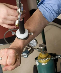
Johns Hopkins researchers are testing an infrared scanning system to detect melanoma (Image: Will Kirk/JHU)
It is responsible for the majority (around 75 percent) of skin cancer related deaths
Although melanoma is one of the less common types of skin cancer, it is responsible for the majority (around 75 percent) of skin cancer related deaths. Part of the problem is that current diagnoses rely on subjective clues such as size, shape and coloring of a mole. With the aim of providing an objective measurement as to whether a lesion may be malignant, researchers at John Hopkins University have developed a prototype non-invasive infrared scanning system that works by looking for the tiny temperature difference between healthy tissue and a growing tumor.
Because cancer cells divide more rapidly than normal cells, they typically generate more metabolic activity and release more energy as heat. Normally, the temperature difference between cancerous and healthy skins cells is extremely small, so Rhoda Alani, adjunct professor at the Johns Hopkins Kimmel Cancer Center and professor and chair of dermatology at the Boston University School of Medicine, teamed with heat transfer expert Cila Herman, a professor of mechanical engineering in Johns Hopkins’ Whiting School of Engineering, to devise a way to make the difference stand out.
Herman aimed her research at measuring heat differences just below the surface of the skin which led to a process that involves first cooling a patient’s skin with a harmless one-minute burst of compressed air. When the cooling is halted, they immediately record infrared images of the target skin area for two to three minutes. Cancer cells typically reheat more quickly than the surrounding healthy tissue, and this difference can be captured by a highly sensitive infrared camera and viewed through sophisticated image processing.
“The system is actually very simple,” Herman said. “An infrared image is similar to the images seen through night-vision goggles. In this medical application, the technology itself is noninvasive; the only inconvenience to the patient is the cooling.”


1 Comments
skincare
melanoma is one of skin cancer type is very difficult to heal.