


Imaging technique tells tumor tissue from normal tissue, could be used in operating room for real-time guidance of surgery
A new laser-based technology may make brain tumor surgery much more accurate, allowing surgeons to tell cancer tissue from normal brain at the microscopic level while they are operating, and avoid leaving behind cells that could spawn a new tumor.
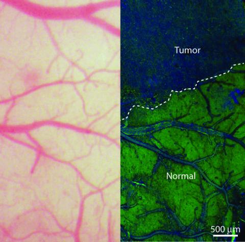
In a new paper, featured on the coverof the journal Science Translational Medicine, a team of University of Michigan Medical School and Harvard University researchers describes how the technique allows them to “see” the tiniest areas of tumor cells in brain tissue.
They used this technique to distinguish tumor from healthy tissue in the brains of living mice — and then showed that the same was possible in tissue removed from a patient with glioblastoma multiforme, one of the most deadly brain tumors.
Now, the team is working to develop the approach, called SRS microscopy, for use during an operation to guide them in removing tissue, and test it in a clinical trial at U-M. The work was funded by the National Institutes of Health.
A need for improvement in tumor removal
On average, patients diagnosed with glioblastoma multiforme live only 18 months after diagnosis. Surgery is one of the most effective treatments for such tumors, but less than a quarter of patients’ operations achieve the best possible results, according to a study published last fall in the Journal of Neurosurgery.
“Though brain tumor surgery has advanced in many ways, survival for many patients is still poor, in part because surgeons can’t be sure that they’ve removed all tumor tissue before the operation is over,” says co-lead author Daniel Orringer, M.D., a lecturer in the U-M Department of Neurosurgery who has worked with the Harvard team since a chance meeting with a team member during his U-M residency.
“We need better tools for visualizing tumor during surgery, and SRS microscopy is highly promising,” he continues. “With SRS we can see something that’s invisible through conventional surgical microscopy.”

The Latest Bing News on:
SRS microscopy
- Challenging Old Theories: Innovative Microscopy Exposes New Alzheimer’s Treatment Pathwayson April 27, 2024 at 3:35 am
Researchers at UC San Diego have utilized advanced imaging techniques to explore the metabolic processes behind Alzheimer's disease, leading to potential new strategies for treatment. Alzheimer's ...
- Innovative Microscopy Demystifies Metabolism of Alzheimer’son April 23, 2024 at 5:00 pm
“Driven by a keen interest in lipid droplet functions in aging and disease, we initiated this fruitful collaboration to harness cutting-edge SRS technology for studying lipid metabolism in tauopathy ...
- Innovative microscopy demystifies metabolism of Alzheimer'son April 23, 2024 at 3:56 pm
Using state-of-the-art microscopy techniques, researchers have shed new light on the underlying mechanisms driving Alzheimer's disease.
- Innovative microscopy demystifies metabolism of Alzheimer’son April 23, 2024 at 9:36 am
“Driven by a keen interest in lipid droplet functions in aging and disease, we initiated this fruitful collaboration to harness cutting-edge SRS technology for studying lipid metabolism in ...
- Innovative microscopy demystifies metabolism of Alzheimer'son April 22, 2024 at 4:59 pm
"Driven by a keen interest in lipid droplet functions in aging and disease, we initiated this fruitful collaboration to harness cutting-edge SRS technology for studying lipid metabolism in ...
- Meibomian Gland Dysfunction: Hyperkeratinization or Atrophy?on April 20, 2024 at 5:00 pm
Probing of tissue sections from normal eyelids using SRS microscopy (c) followed by staining for actin (d, Phalloidin) and nuclei (d, DAPI) showed lipid signals within the gland that appeared ...
- Advanced microscopy technique offers a new look inside cellson April 10, 2024 at 5:00 pm
Researchers face a similar dilemma when imaging cells under a microscope. Within a single cell lives an intricate ecosystem of millions of molecules interacting with one another. Viewing ...
- Laser tricks without labelson March 10, 2024 at 6:50 pm
elegans to probe thousands of genes for their role in lipidogenesis and using a microscopy technique called stimulated Raman scattering (SRS) to monitor the results. Ruvkun's collaborator is ...
- Understanding Scanning Tunneling Microscopy for Precise Surface Analysison February 16, 2024 at 2:30 pm
Scanning Tunneling Microscopy is a subset of Scanning Probe Microscopy (SPM), a group of techniques that image and manipulate surfaces at the nanometer to atomic scales. SPM techniques share the ...
- What You Need To Know About Microplastics In Bottled Wateron January 27, 2024 at 7:13 am
It's common knowledge at this point that plastic doesn't break down the way paper products do. During its tenure in a landfill, rather than decompose, plastic breaks down into smaller and smaller ...
The Latest Google Headlines on:
SRS microscopy
[google_news title=”” keyword=”SRS microscopy” num_posts=”10″ blurb_length=”0″ show_thumb=”left”]
The Latest Bing News on:
Brain tumor surgery
- KOTA Cares: Rapid City 2nd grader battles rare brain tumoron May 9, 2024 at 5:00 am
For this week’s KOTA Cares, we spotlight one little girl in Rapid City who could use all the support she can get while battling a rare brain tumor. After undergoing surgery at the Mayo Clinic in the spring of 2022,
- Isabella Strahan Gives Brain Cancer Treatment Update After Unexpected Surgeryon May 8, 2024 at 1:06 pm
Isabella Strahan shared a health update amid her ongoing journey with brain cancer. Her latest vlog shares a few days at home after being hospitalized in late March and early April. She is back to resting and regular appointments after undergoing unexpected brain surgery.
The Latest Google Headlines on:
Brain tumor surgery
[google_news title=”” keyword=”brain tumor surgery” num_posts=”10″ blurb_length=”0″ show_thumb=”left”]

