


Super-sharp images from within the human body made through tiny endoscopes have come a step closer to reality thanks to joint research by scientists from the University of Twente’s MESA+ research institute, the Max Planck Institute for the Science of Light (MPL), FOM and Carl Zeiss AG.
An advanced wavefront shaping method developed at the UT combined with unique optical fibres from MPL make it possible to focus lensless light at an unparalleled resolution. FOM postdoc Dr Lyubov Amitonova and her colleagues published their findings on 29 January in Optics Letters, the leading journal of the Optical Society of America.
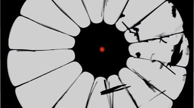
Optical imaging via ultrathin fibre probes is extremely useful for taking a look inside the human body in a minimally invasive manner. Unfortunately, the resolution of current fibre endoscopes is one micrometre at best. This is not enough to see interesting and important fine features in biological cells, for example. Some endoscopes use a high number of separate fibres bound together into a fibre bundle. Each fibre then acts like a discrete pixel to form the final pixelated image. However such bundles tend to be quite thick, at least a millimetre in diameter.
An alternative is fibre endoscopes based on ‘multimode’ fibres. These could offer imaging with a better view and be as thin as a tenth of a millimetre. A multimode fibre uses only a single fibre core that can transmit an entire image. Unfortunately, the image becomes scrambled as it passes through the fibre. However, some tricks for unscrambling these images are available. The main limiting factor for the resolution of these multimode endoscopes is that the fibres only transmit light that propagates along the fibre axis. Light under a small angle can still bounce from the fibre walls. But if this angle gets too large, the light will simply leak out to the side. FOM postdoc Dr Lyubov Amitonova and her colleagues at UT and MPL have now shown that with photonic crystal fibres this limitation can be overcome.
UNIQUE CRYSTAL FIBRE PROBE
Conventional (‘step-index’) fibres consist of two zones of different material (an outer cladding and an inner core) with distinct refraction indices, which enable light transmission down the fibre axis by total internal reflection. Photonic crystal fibres are built differently: they are made of one material only and light guiding is realised through the presence of a specific pattern of holes in the cladding, which are filled with air. Tailoring the cladding structure of such a fibre provides a unique tool for engineering specific fibre-optic properties. In this project, the scientists have designed and made such a fibre to focus a laser beam through the fibre down to 0.52 micrometres, using visible red light.
SHARP FOCUS AND HIGH RESOLUTION
A photonic crystal fibre acts as a multimode fibre in which the image typically gets scrambled due to light bouncing off the possibly irregular wall of the fibre. The technique of complex wavefront shaping, invented at UT, is able to undo such scrambling and make a sharp focus. This is achieved by pre-shaping the light into the precise form needed to make a sharp image behind the fibre before the light actually enters the fibre. Using this approach, Amitonova and her colleagues have succeeded in focusing light at the fibre output facet of different multimode fibres including several unique photonic crystal fibres. They have shown that the complex wavefront shaping technique together with a properly designed multimode photonic crystal fibre allows the creation of a tightly focused spot at the desired position on the fibre output facet with a subwavelength beam waist.
This paves the way towards high-resolution endoscopic imaging via fibre probes so thin that they could be inserted, for instance, into tiny blood vessels not much thicker than a human hair.
Read more: Super-sharp images through thin optical fibres
The Latest on: High-resolution endoscopic imaging
[google_news title=”” keyword=”high-resolution endoscopic imaging” num_posts=”10″ blurb_length=”0″ show_thumb=”left”]
via Google News
The Latest on: High-resolution endoscopic imaging
- Optical barcodes expand range of high-resolution sensoron April 26, 2024 at 11:27 am
The same geometric quirk that lets visitors murmur messages around the circular dome of the whispering gallery at St. Paul's Cathedral in London or across St. Louis Union Station's whispering arch ...
- GI & Endoscopy E-Newsletteron April 26, 2024 at 9:23 am
To sign up for Becker's GI & Endoscopy E-Newsletter or any of our other E-Newsletters, click here. If you are experiencing difficulty receiving our newsletters, you may need to whitelist our new ...
- Advancing high-resolution ultrasound imaging with deep learningon April 22, 2024 at 11:38 am
Researchers at the Beckman Institute for Advanced Science and Technology have developed a new technique to make ultrasound localization microscopy, an emerging diagnostic tool used for high-resolution ...
- Diagnosis of Undifferentiated Type Early Gastric Cancers by Magnification Endoscopy With Narrow-band Imagingon April 17, 2024 at 5:00 pm
All endoscopic examinations were performed using either a high-resolution magnification upper ... both low-magnification and high-magnification imaging in all of the consecutive cases.
- Researchers develop 3D imaging probe for cancer cell detectionon April 16, 2024 at 10:49 am
A research team at the University of Nottingham has developed an endoscopic probe enabling practitioners to three-dimensionally (3D) image the stiffness of individual biological cells and complex ...
- World-first microscopic stiffness probe could advance early cancer diagnosison April 15, 2024 at 8:17 am
Researchers at the University of Nottingham have created an endoscopic device that can 3D image the stiffness of individual biological cells and complex organisms, a discovery that could help doctors ...
- Fujifilm Receives 510(k) Clearance for CAD EYE®, New AI-Powered Endoscopic Imaging Technology for Colonic Polyp Detectionon March 20, 2024 at 2:05 am
Lexington, Mass., March 20, 2024 (GLOBE NEWSWIRE) -- FUJIFILM Healthcare Americas Corporation, a leading provider of endoscopic imaging and endosurgical solutions, received 510(k) clearance for CA ...
- Insulinoma—new insights into an old diseaseon December 26, 2023 at 8:38 am
A combination of high-resolution imaging techniques, endoscopic ultrasonography, angiography with selective arterial-calcium stimulation test and intraoperative ultrasonography is of paramount ...
- The power to see nanoscale structures inside cellson August 10, 2022 at 6:37 am
Endoscopy on ... over other nano-imaging techniques, such as cryogenic electron microscopy and super-resolution fluorescent microscopy, Fukuma notes. One is its high spatial resolution.
- High Resolution Brain Imaging Labon December 18, 2020 at 3:49 am
In our lab, we develop and utilize sophisticated magnetic resonance imaging (MRI) methods to study a variety ... to human sensory perception and attention, addressed using high-resolution MRI methods ...
via Bing News
