


Purdue University researchers are developing a novel biomedical imaging system that combines optical and ultrasound technology to improve diagnosis of life-threatening diseases.
Photoacoustic tomography is a noninvasive technique that works by converting absorbed optical energy into acoustic signal. Pulsed light is sent into body tissue, creating a small increase in temperature that causes the tissue to expand and create an acoustic response that can be detected by an ultrasound transducer. The ultrasound data is used to visualize the tissue.
“The nice thing about photoacoustic tomography is the compositional information,” said Craig Goergen, an assistant professor in Purdue’s Weldon School of Biomedical Engineering. “It provides information about where blood and lipid are located, along with other essential information.”
The ultimate goal is to enhance the clinical care of patients.
The results of a study describing an adjustable photoacoustic probe with improved light delivery and image quality were published Tuesday (Aug. 28) in the journal Photoacoustics.
The system provides real-time compositional information of body tissue without the need for contrast agents and with better depth penetration compared with conventional optical techniques.
Photoacoustic tomography can be used to detect or monitor a myriad of diseases, including cardiovascular disease, diabetes, and cancer. Those are diseases that the Centers for Disease Control and Prevention lists as among the most common, costly, and preventable of all health problems. Heart disease and cancer each account for one in every four deaths a year in the United States, and more than 30 million Americans, or more than 9 percent of the population, have diabetes. The cost of those three diseases a year in the United States is more than $718 billion a year, according to the CDC.
“That means there will be a great need for medical imaging. Trying to diagnose these diseases at an earlier time can lead to improved patient care,” Goergen said. “We are in the process now of trying to use this enhanced imaging approach to a variety of different applications to see what it can be used for.”
Among other potential uses for photoacoustic tomography is the mapping of lipid deposition within an arterial wall that can cause other health problems, measuring cardiac tissue damage and tumor biopsies. Using photoacoustic tomography for intraoperative tumor biopsies could help surgeons make sure they remove all the cancer from a patient, Goergen said.
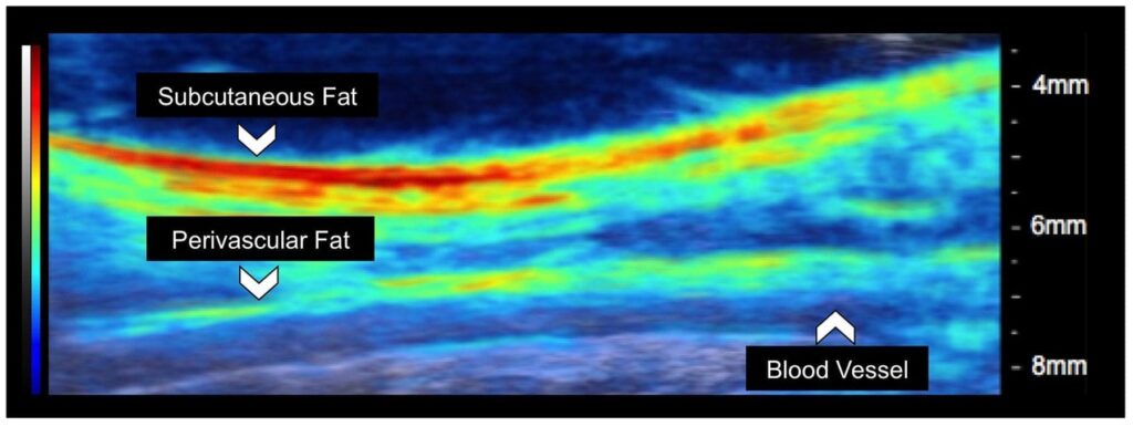
One of the challenges of photoacoustic tomography is improving the penetration depth and signal-to-noise ratio past optical absorbers. The researchers believe creating optical manipulation techniques to maximize photon density could provide a solution. As a result, they have created a motorized photoacoustic holder that allows users to easily maneuver the aim of the device and tune the depth where light is focused, improving the light penetration depth and signal-to-noise ratio.
A video about the acoustic tomography is available at https://bit.ly/2yJddb0. A complete list of co-authors is available in the abstract. The research has been funded by the National Institutes of Health.
The Purdue researchers are interested in talking with other companies about other possible uses for photoacoustic tomography. The researchers have a patent pending for the technology with the help of the Purdue Office of Technology Commercialization.
Learn more: Purdue researchers developing novel biomedical imaging system combining optical, ultrasound technology
The Latest on: Biomedical imaging
[google_news title=”” keyword=”biomedical imaging” num_posts=”10″ blurb_length=”0″ show_thumb=”left”]
via Google News
The Latest on: Biomedical imaging
- New imaging technique provides a much closer look at fibril assemblieson April 26, 2024 at 10:47 pm
A new imaging technique developed by engineers at Washington University in St. Louis can give scientists a much closer look at fibril assemblies, stacks of peptides like amyloid beta, most notably ...
- Library Faculty to Help Researchers Access Massive Biomedical Databaseon April 25, 2024 at 11:28 am
UM Libraries received the $40,000 All of Us Academic Libraries Program award. Library faculty Savannah Kelly and Shelby Watson will offer workshops for both beginning and veteran researchers on how to ...
- Perinatal substance use may shape how strongly mothers feel toward infantson April 25, 2024 at 10:22 am
Substance use during pregnancy and postpartum may impact areas of the brain associated with what psychologists and neuroscientists call "affective empathy," or the emotional response experienced as a ...
- Biomedical Engineer Salaryon April 24, 2024 at 5:00 pm
How Much Does a Biomedical Engineer Make? Biomedical Engineers made a median salary of $99,550 in 2022. The best-paid 25% made $129,230 that year, while the lowest-paid 25% made $78,500.
- Imaging technique shows new details of peptide structureson April 24, 2024 at 11:19 am
A new imaging technique developed by engineers at Washington University in St. Louis can give scientists a much closer look at fibril assemblies—stacks of peptides that include amyloid beta, most ...
- Biomedical Applications of Nanodiamonds in Imaging and Therapyon April 19, 2024 at 4:59 pm
In this review an attempt is made to capture the scope, spirit and recent developments in the field of nanodiamonds for biomedical applications ... optimizing them for imaging and sensing ...
- Researchers review use of MRI to identify brain cancer biomarkerson April 11, 2024 at 12:15 pm
Researchers from the School of Biomedical Engineering & Imaging Sciences (BMEIS) have published a systematic review in Neuro-Oncology Advances exploring the use of MRI imaging techniques to identify ...
- Biomedical Applications of Nanodiamonds in Imaging and Therapyon April 7, 2024 at 5:00 pm
In addition to the biomedical applications of NDs in the ... the spectroscopic properties for adjusting/optimizing them for imaging and sensing applications are still under investigation.
- Updated with memorial fund information: Biomedical Engineering Chair Joseph Izatt Dieson April 7, 2024 at 5:00 pm
The lab’s expertise in OCT technology has also allowed them to expand their reach beyond the realm of biomedical imaging to other endeavors, such as investigating how OCT could help autonomous robots ...
- Data-driven polarimetric imagingon April 3, 2024 at 5:00 pm
Existing data-driven polarization imaging technologies have gradually found applications in polarization information reconstruction and enhancement, target detection, biomedical imaging ...
via Bing News