


via T. Lasser/EPFL
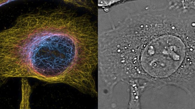
Super resolution microscopy is a technique that can see beyond the diffraction of light and provides unprecedented views of cells and their interior structures and organelles.
An EPFL team has developed a technique that can perform both 3D super resolution microscopy and fast 3D phase imaging in a single instrument.
This unique 4D microscope combines a sensitivity and high time resolution of phase imaging with the specifically and high spatial resolution of fluorescence microscopy. This allows the direct visualization and Analysis of subcellular structures in living cells without labeling.
These technical advances enable high-resolution visualization of the formation of pathological alpha-synuclein aggregates in hippocampal neurons. It is hoped that it will become a regular workhorse for neuroscience and biology.
Learn more: Super-resolution microscopy in both space and time
The Latest on: Super resolution microscopy
[google_news title=”” keyword=”Super resolution microscopy” num_posts=”10″ blurb_length=”0″ show_thumb=”left”]
via Google News
The Latest on: Super resolution microscopy
- Feed has no items.
via Bing News

