


Researchers have found a way to see synthetic nanostructures and molecules using a new type of super-resolution optical microscopy that does not require fluorescent dyes, representing a practical tool for biomedical and nanotechnology research.
“Super-resolution optical microscopy has opened a new window into the nanoscopic world,” said Ji-Xin Cheng, an associate professor of biomedical engineering and chemistry at Purdue University.
Conventional optical microscopes can resolve objects no smaller than about 300 nanometers, or billionths of a meter, a restriction known as the “diffraction limit,” which is defined as half the width of the wavelength of light being used to view the specimen. However, researchers want to view molecules such as proteins and lipids, as well as synthetic nanostructures like nanotubes, which are a few nanometers in diameter.
Such a capability could bring advances in a diverse range of disciplines, from medicine to nanoelectronics, Cheng said.
“The diffraction limit represents the fundamental limit of optical imaging resolution,” Cheng said. “Stefan Hell at the Max Planck Institute and others have developed super-resolution imaging methods that require fluorescent labels. Here, we demonstrate a new scheme for breaking the diffraction limit in optical imaging of non-fluorescent species. Because it is label-free, the signal is directly from the object so that we can learn more about the nanostructure.”
Findings are detailed in a research paper that appeared online Sunday (April 28) in the journal Nature Photonics.
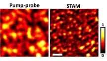
The imaging system, called saturated transient absorption microscopy, or STAM, uses a trio of laser beams, including a doughnut-shaped laser beam that selectively illuminates some molecules but not others. Electrons in the atoms of illuminated molecules are kicked temporarily into a higher energy level and are said to be excited, while the others remain in their “ground state.” Images are generated using a laser called a probe to compare the contrast between the excited and ground-state molecules.
The researchers demonstrated the technique, taking images of graphite “nanoplatelets” about 100 nanometers wide.
“It’s a proof of concept and has great potential for the study of nanomaterials, both natural and synthetic,” Cheng said.
The Latest Bing News on:
Super-resolution optical microscopy
- Super-resolution microscopy articles from across Nature Portfolioon April 23, 2024 at 5:00 pm
Super-resolution microscopy includes a variety ... control of the switching of fluorophores in stochastic optical reconstruction microscopy (EC-STORM) enables the counting of single fluorophores ...
- STORM Microscopy: Single-Molecule Localization for Nanoscale Imaging of Biological Structureson April 15, 2024 at 1:09 pm
Stochastic Optical Reconstruction Microscopy (STORM) is a super-resolution imaging technique that allows researchers to visualize biological structures and processes at the nanoscale. It overcomes the ...
- Super-Resolution Microscopy Can Be Super-Accessibleon April 8, 2024 at 5:00 pm
A decade ago, the Nobel Prize in Chemistry was awarded to a trio of researchers for the development of super-resolved fluorescence microscopy. The announcement at the time stated that the ...
- Artificial intelligence boosts super-resolution microscopyon March 28, 2024 at 10:45 am
Super-resolution microscopy aims to overcome the diffraction limit, a limit that restricts resolution due to the optical characteristics of the microscopic system. To surmount this limit ...
- Vutara VXL-Super-resolution microscopy workstationon July 17, 2023 at 7:10 am
Several fascinating cellular structures are smaller than the optical diffraction limit of approximately 200 to 300 nm and hence need super-resolution microscopy for imaging specifically labeled ...
- Eyes on super-resolutionon April 29, 2023 at 5:23 pm
The outlook is truly exciting. As more and more molecules and methods for super-resolution imaging are developed, the power of optical microscopy to observe complex nanoscale systems non ...
- Super-Resolution STORMon March 24, 2021 at 7:27 pm
Stochastic Optical Reconstruction Microscopy (STORM) reconstructs a super-resolution fluorescence image by combining precise localization information for individual fluorophores in complex fluorescent ...
- Super-Resolution Microscope Functionality for EMCCD - iXon SRRF-Streamon August 14, 2020 at 6:19 pm
100 raw ‘input’ images were recorded for every resultant super-resolution image ... recorded on the same microscope, using identical objective and optical path. The sole difference being that SIM was ...
- Super-resolution microscopyon October 17, 2017 at 6:17 am
STED (Stimulated emission depletion) is a true optical super-resolution technique in which a doughnut shaped ... TIRF imaging by our Abbelight SAFe360 system on Olympus IX83 microscope.
The Latest Google Headlines on:
Super-resolution optical microscopy
[google_news title=”” keyword=”super-resolution optical microscopy” num_posts=”10″ blurb_length=”0″ show_thumb=”left”]
The Latest Bing News on:
Super-resolution microscope
- Purdue-created technology makes 3D microscopes easier to use, less expensive to manufactureon April 30, 2024 at 11:10 am
Liming Chen, who will earn his PhD in mechanical engineering from Purdue in May 2024, operates a 3D microscope, which uses an optical technique called fringe projection to create a high-resolution 3D ...
- Imaging Technique Shows New Details of Alzheimer’s Peptide Structureson April 29, 2024 at 1:38 am
A new imaging technique developed by engineers at Washington University in St. Louis can give scientists a much closer look at fibril assemblies — stacks of peptides that include amyloid beta, most ...
- New imaging technique provides a much closer look at fibril assemblieson April 26, 2024 at 10:47 pm
A new imaging technique developed by engineers at Washington University in St. Louis can give scientists a much closer look at fibril assemblies, stacks of peptides like amyloid beta, most notably ...
- Imaging technique shows new details of peptide structureson April 25, 2024 at 5:00 pm
Researchers outline how they used a chemical probe to light up interlocking peptides. Their technique will help scientists differentiate synthetic peptides from toxic types found in Alzheimer's ...
- Testing how well biomarkers work: New fluorescence microscopy method can improve resolution down to the Ångström scaleon April 24, 2024 at 9:49 am
LMU researchers have developed a method to determine how reliably target proteins can be labeled using super-resolution fluorescence microscopy.
- 30 Times Clearer – Scientists Develop Improved Mid-Infrared Microscopeon April 24, 2024 at 3:55 am
The chemical images taken of the insides of bacteria were 30 times clearer than those from conventional mid-infrared microscopes. Researchers at the University of Tokyo have developed an advanced ...
- Scientists Solve Decades-Old Microscopy Problemon April 24, 2024 at 1:55 am
Studying tissues, cells, and proteins under a microscope is essential for disease prevention and treatment. This research requires accurately measuring the dimensions of these biological structures.
- Biophysics: Testing how well biomarkers workon April 23, 2024 at 5:00 pm
Researchers have developed a method to determine how reliably target proteins can be labeled using super-resolution fluorescence microscopy. LMU researchers have developed a method to determine how ...
- World’s only quantum-gas microscope imaging strontium’s individual atomson April 23, 2024 at 4:40 am
Spanish researchers develop the world's first quantum-gas microscope that captures images of individual atoms of strontium quantum gases.
- New super-resolution microscopy approach visualizes internal cell structures and clusters via selective plane activationon April 22, 2024 at 9:08 am
To study living organisms at ever smaller length scales, scientists must devise new techniques to overcome the so-called diffraction limit. This is the intrinsic limitation on a microscope's ability ...
The Latest Google Headlines on:
Super-resolution microscope
[google_news title=”” keyword=”super-resolution microscope” num_posts=”10″ blurb_length=”0″ show_thumb=”left”]

