


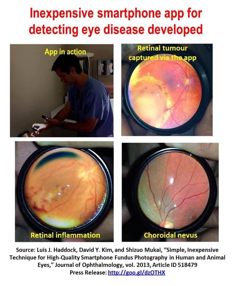
Retinal (or fundus) photography is an essential part of any ophthalmology practice.
Commercial fundus cameras can cost tens to hundreds of thousands of dollars, making the technology out of reach for smaller ophthalmic practices and to physicians in third-world countries. In a recent study now on line, Massachusetts Eye and Ear researchers describe the relatively simple technique of fundus photography in human and rabbit eyes using a smartphone, an inexpensive app for the smartphone, and instruments that are readily available in an ophthalmic practice.
Smartphones are now being used more routinely in ophthalmology to document patients’ ocular conditions, the authors write. Previously described techniques of fundus imaging often proved difficult to repeat, partly because video capture using Apple’s built-in camera app in the iPhones cannot independently control the focus and the exposure during filming, which results in glare and poor image quality.
“Our technique provides a simpler and higher quality method to more consistently produce excellent images of a patient’s fundus,” said senior author Shizuo Mukai, M.D.,Mass. Eye and Ear retina specialist and Harvard Medical School associate professor of Ophthalmology. “This technique has been extremely helpful for us in the emergency department setting, in-patient consultations, and during examinations under anesthesia as it provides a cheaper and portable option for high-quality fundus-image acquisition for documentation and consultation. This technique is well tolerated in awake patients most likely since the light intensity used is often well below that which is used in standard indirect ophthalmoscopy.”
Using the described technique of smartphone fundus photography with the use of iPhone 4 or iPhone 5, the app Filmic pro, and a 20D lens with or without a Koeppe lens, researchers were was able to capture excellent, high-quality fundus images in both children under anesthesia and in awake adults.
The Latest Bing News on:
Diagnosing eye disease
- What Is Eye Syphilis? The Severe Symptom Doctors Are Seeing amid the STD Epidemicon April 26, 2024 at 2:46 pm
Doctors are seeing more patients with unusual vision and eye symptoms due to the sexually transmitted infection ...
- Embiid's diagnosis draws new attention to Bell's palsyon April 26, 2024 at 2:15 pm
Philadelphia 76ers All-Star center Joel Embiid has been diagnosed with Bell's palsy. A facial nerve gets inflamed or injured and suddenly muscles on one side of the face become weak or paralyzed.
- Missy Elliott's Graves' Disease Caused 'Extreme' Weight Loss: Here's What She's Shared About Her Conditionon April 26, 2024 at 9:41 am
Symptoms include weight loss (despite having an increased appetite), a rapid or irregular heartbeat, fatigue, shaky hands, and trouble tolerating heat, the NIDDK says. Missy revealed that she was ...
- Imaging using fundus autofluorescence can facilitate the diagnosis and monitoring of rare eye diseaseson April 25, 2024 at 9:27 pm
Uveitis is a rare inflammatory eye disease. Posterior and panuveitis in particular are associated with a poor prognosis and a protracted course of the disease. Diagnosis and monitoring can be ...
- Diagnosis of rare eye diseases: Uveitis experts provide an overview of an underestimated imaging techniqueon April 25, 2024 at 6:51 am
Uveitis is a rare inflammatory eye disease. Posterior and panuveitis in particular are associated with a poor prognosis and a protracted course of the disease. Diagnosis and monitoring can be ...
- Simplified diagnosis of rare eye diseaseson April 25, 2024 at 6:43 am
Uveitis is a rare inflammatory disease of the choroid of the eye, which lies between the retina ... posterior and panuveitis. The exact diagnosis of posterior uveitis and panuveitis can be ...
- Simplified diagnosis of rare eye diseases - Uveitis experts provide an overview of an underestimated imaging techniqueon April 25, 2024 at 2:26 am
Mail: [email protected] Matthias M. Mauschitz, Markus Zeller et al: Fundus Autofluorescence in Posterior and Panuveitis - An Under-Estimated Imaging Technique: A Review and Case Series; ...
- AI tool recognizes serious ocular disease in horseson April 24, 2024 at 1:02 pm
Researchers have developed a deep learning tool that is capable of reliably diagnosing moon blindness in horses based on photos.
- AI-Powered Solutions for Personalized Therapeutic Interventions in Dry Eye Diseaseon April 24, 2024 at 12:44 pm
According to a study published in Big Data Mining and Analytics, researchers want to employ artificial intelligence (AI) to help in the early detection and diagnosis of Dry Eye Disease (DED).
- Could a simple eye test predict Alzheimer's 12 years before symptoms show?on April 18, 2024 at 6:00 am
A recent study suggests that people with poor vision sensitivity are more likely to go on and develop Alzheimer's disease, and that a simple eye test could help predict the onset of this condition.
The Latest Google Headlines on:
Diagnosing eye disease
[google_news title=”” keyword=”diagnosing eye disease” num_posts=”10″ blurb_length=”0″ show_thumb=”left”]
The Latest Bing News on:
Fundus photography
- Imaging using fundus autofluorescence can facilitate the diagnosis and monitoring of rare eye diseaseson April 25, 2024 at 9:27 pm
Uveitis is a rare inflammatory eye disease. Posterior and panuveitis in particular are associated with a poor prognosis and a protracted course of the disease. Diagnosis and monitoring can be ...
- Diagnosis of rare eye diseases: Uveitis experts provide an overview of an underestimated imaging techniqueon April 25, 2024 at 6:51 am
Uveitis is a rare inflammatory eye disease. Posterior and panuveitis in particular are associated with a poor prognosis and a protracted course of the disease. Diagnosis and monitoring can be ...
- Photography Minoron April 7, 2024 at 5:00 pm
The photography minor explores the diverse subject of photography from either an art or science perspective. Students develop both technical and aesthetic skills needed for creative, communication, or ...
- Optical Coherence Tomography and Fundus Autofluorescence Imaging in Uveitison March 2, 2024 at 4:00 pm
Fundus autofluorescence (FAF) is an imaging technique that is used to evaluate retinal disorders that affect the retinal pigment epithelium (RPE). FAF is useful to evalute disease activity in a ...
- Diabetic Retinopathy Management Guidelineson December 14, 2023 at 8:23 pm
optometrists and other trained medical examiners Canada Seven-standard field stereoscopic colour fundus photography or dilated direct ophthalmoscopy or dilated indirect slit-lamp fundoscopy Fundus ...
- Travel Photography Tipson August 15, 2020 at 6:12 pm
Serendipity plays an enormously important role in travel photography. You never know what you are going to run into, and you have to be ready. Many times you will see what could be a good ...
- Action and Adventure Photography Tipson March 3, 2015 at 9:02 pm
In a large part, adventure photography is about telling a story. I always use a story line in a trip. I start this process at the beginning of a trip, and I become more intimately involved and ...
The Latest Google Headlines on:
Fundus photography
[google_news title=”” keyword=”fundus photography” num_posts=”10″ blurb_length=”0″ show_thumb=”left”]
