


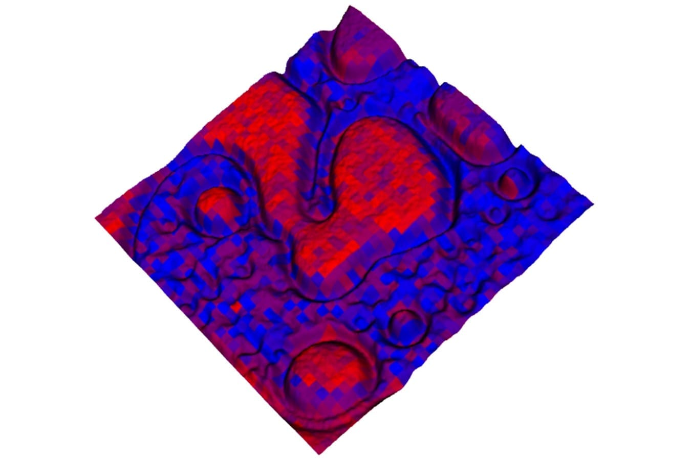
A tool that provides world-class microscopy and spatially resolved chemical analysis shows considerable promise for advancing a number of areas of study, including chemical science, pharmaceutical development and disease progression.
The hybrid optical microscope/mass spectrometry-based imaging system developed at the Department of Energy’s Oak Ridge National Laboratory operates under ambient conditions and requires no pretreatment of samples to analyze chemical compounds with sub-micron resolution. One micron is equal to about 1/100th the width of a human hair. Results of the work by postdoctoral associate Jack Cahill and Gary Van Berkel and Vilmos Kertesz of ORNL’s Chemical Sciences Division are detailed in Analytical Chemistry.
“Knowing the chemical basis of material interactions that take place at interfaces is vital for designing and advancing new functional materials that are important for DOE missions such as organic photovoltaics for solar energy,” Van Berkel said. “In addition, the new tool can be used to better understand the chemical basis of important biological processes such as drug transport, disease progression and response for treatment.”
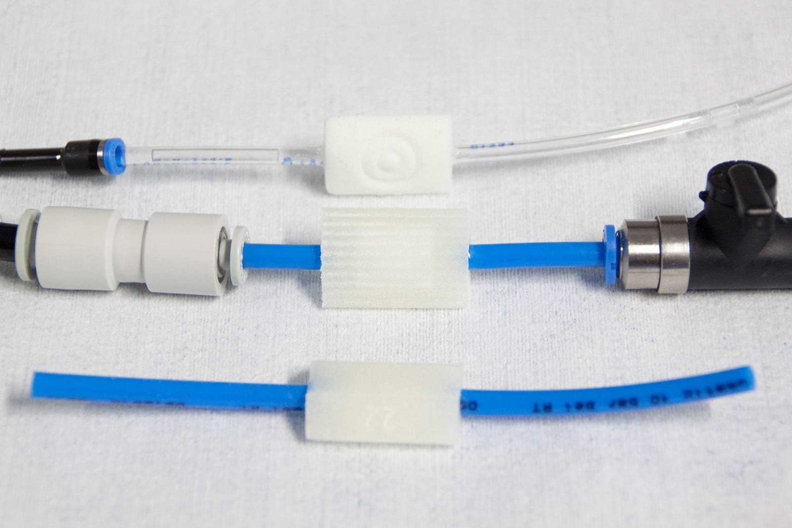
The hybrid instrument transfers tiny amounts of a material such as human tissue or an organic polymer from a sample by a laser ablation process in which material is captured and transported via liquid stream to the ionization source of the mass spectrometer. In just seconds, a computer screen displays the results.
Researchers noted that the resolution of less than one micron is essential to accurately differentiate and distinguish between polymers and sub-components of similar-sized cells.
“Today’s mass spectrometry imaging techniques are not yet up to the task of reliably acquiring molecular information on a wide range of compound types,” Cahill said. “Examples include synthetic polymers used in various functional materials like light harvesting and emitting devices or biopolymers like cellulose in plants or proteins in animal tissue.”
This technology, however, provides the long-sought detailed chemical analysis through a simple interface between a hybrid optical microscope and an electrospray ionization system for mass spectrometry.
Read more: New ORNL device combines power of mass spectrometry, microscopy
The Latest on: Microscopy
[google_news title=”” keyword=”microscopy” num_posts=”10″ blurb_length=”0″ show_thumb=”left”]
via Google News
The Latest on: Microscopy
- Challenging Old Theories: Innovative Microscopy Exposes New Alzheimer’s Treatment Pathwayson April 27, 2024 at 3:35 am
Researchers at UC San Diego have utilized advanced imaging techniques to explore the metabolic processes behind Alzheimer's disease, leading to potential new strategies for treatment. Alzheimer's ...
- ZEISS and Argolight announce partnership to enhance microscopy imaging quality controlon April 24, 2024 at 6:01 pm
ZEISS and Argolight announced their strategic partnership. The collaboration aims to seamlessly integrate cutting-edge quality control solutions of the French company into the extensive line of ZEISS ...
- One-of-a-kind science facility in Stockton trains next generation for high-paying careerson April 24, 2024 at 5:57 pm
Delta College's electron microscopy program is the only one of its kind in the nation at a community college level.
- Scientists pioneer new X-ray microscopy method for data analysis 'on the fly'on April 24, 2024 at 9:54 am
A new streaming technique allows playback of data while it is being generated. When scientists want to look at a tiny structure in a material, even one just a few atoms in size, they frequently turn ...
- Testing how well biomarkers work: New fluorescence microscopy method can improve resolution down to the Ångström scaleon April 24, 2024 at 9:49 am
LMU researchers have developed a method to determine how reliably target proteins can be labeled using super-resolution fluorescence microscopy.
- Scientists Solve Decades-Old Microscopy Problemon April 24, 2024 at 1:55 am
Studying tissues, cells, and proteins under a microscope is essential for disease prevention and treatment. This research requires accurately measuring the dimensions of these biological structures.
- Biophysics: Testing how well biomarkers workon April 23, 2024 at 5:00 pm
Researchers have developed a method to determine how reliably target proteins can be labeled using super-resolution fluorescence microscopy. LMU researchers have developed a method to determine how ...
- Innovative microscopy demystifies metabolism of Alzheimer'son April 23, 2024 at 3:56 pm
Using state-of-the-art microscopy techniques, researchers have shed new light on the underlying mechanisms driving Alzheimer's disease.
- Open house at Delta showcases only community college electron microscopy program in the USAon April 19, 2024 at 1:29 am
Discover worlds within our world as Delta College hosts a free open house on Tuesday, April 23. showcasing the College’s unique electron microscopy program.
- Microscopy News and Researchon April 16, 2024 at 5:01 pm
This is the first time that all the proteins of an organism have been tracked across the cell cycle, which required a combination of deep learning and high-throughput microscopy. Low birth rates ...
via Bing News
