


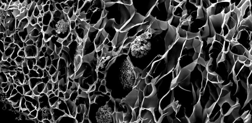
A tiny and intricate biomedical structure created with the new technique.
Researchers have flipped traditional 3D printing to create some of the most intricate biomedical structures yet, advancing the development of new technologies for regrowing bones and tissue.
The emerging field of tissue engineering aims to harness the human body’s natural ability to heal itself, to rebuild bone and muscle lost to tumours or injuries.
A key focus for biomedical engineers has been the design and development of 3D printed scaffolds that can be implanted in the body to support cell regrowth.
But making these structures small and complex enough for cells to thrive remains a significant challenge.
Enter a RMIT University-led research team, collaborating with clinicians at St Vincent’s Hospital Melbourne, who have overturned the conventional 3D printing approach.
Instead of making the bioscaffolds directly, the team 3D printed moulds with intricately-patterned cavities then filled them with biocompatible materials, before dissolving the moulds away.
Using the indirect approach, the team created fingernail-sized bioscaffolds full of elaborate structures that, until now, were considered impossible with standard 3D printers.
Lead researcher Dr Cathal O’Connell said the new biofabrication method was cost-effective and easily scalable because it relied on widely available technology.
“The shapes you can make with a standard 3D printer are constrained by the size of the printing nozzle – the opening needs to be big enough to let material through and ultimately that influences how small you can print,” O’Connell, a Vice-Chancellor’s Postdoctoral Fellow at RMIT, said.
“But the gaps in between the printed material can be way smaller, and far more intricate.
“By flipping our thinking, we essentially draw the structure we want in the empty space inside our 3D printed mould. This allows us to create the tiny, complex microstructures where cells will flourish.”
Versatile technique
O’Connell said other approaches were able to create impressive structures, but only with precisely-tailored materials, tuned with particular additives or modified with special chemistry.
“Importantly, our technique is versatile enough to use medical grade materials off-the-shelf,” he said.
“It’s extraordinary to create such complex shapes using a basic ‘high school’ grade 3D printer.
“That really lowers the bar for entry into the field, and brings us a significant step closer to making tissue engineering a medical reality.”
The research, published in Advanced Materials Technologies, was conducted at BioFab3D@ACMD, a state-of-the-art bioengineering research, education and training hub located at St Vincent’s Hospital Melbourne.
Co-author Associate Professor Claudia Di Bella, an orthopedic surgeon at St Vincent’s Hospital Melbourne, said the study showcases the possibilities that open up when clinicians, engineers and biomedical scientists come together to address a clinical problem.
“A common problem faced by clinicians is the inability to access technological experimental solutions for the problems they face daily,” Di Bella said.
“While a clinician is the best professional to recognise a problem and think about potential solutions, biomedical engineers can turn that idea into reality.
“Learning how to speak a common language across engineering and medicine is often an initial barrier, but once this is overcome, the possibilities are endless.”
Di Bella said clinicians, like surgeons, are used to applying their manual skills, as well as technical tools, to “fix” a problem within the body.
“Bioengineering technologies can significantly increase the armoury of options available to clinicians and offer the possibility to address problems never solved before, as well as to give personalised, patient-specific, solutions.
“This is an incredible step forward in medicine and one that excites patients and clinicians alike.”
Future treatment toolkit
Currently there are few treatment options for people who lose a significant amount of bone or tissue due to illness or injury, making amputation or metal implants to fill a gap common outcomes.
While a few clinical trials of tissue engineering have been conducted around the world, key bioengineering challenges still need to be addressed for 3D bioprinting technology to become a standard part of a surgeon’s toolkit.
In orthopedics, a major sticking point is the development of a bioscaffold that works across both bone and cartilage.
“Our new method is so precise we’re creating specialised bone and cartilage-growing microstructures in a single bioscaffold,” O’Connell said.
“It’s the surgical ideal – one integrated scaffold that can support both types of cells, to better replicate the way the body works.”
Tests with human cells have shown bioscaffolds built using the new method are safe and non-toxic.
The next steps for the researchers will be testing designs to optimise cell regeneration and investigating the impact on cell regrowth of different combinations of biocompatible materials.
Step-by-step: How to reverse print a bioscaffold
The new method – which researchers have dubbed Negative Embodied Sacrificial Template 3D (NEST3D) printing – uses simple PVA glue as the basis for the 3D printed mould.
Once the biocompatible material injected into the mould has set, the entire structure is placed in water to dissolve the glue, leaving just the cell-nurturing bioscaffold.
Study first author, PhD researcher Stephanie Doyle, said the method enabled researchers to rapidly test combinations of materials to identify those most effective for cell growth.
“The advantage of our advanced injection moulding technique is its versatility,” Doyle said.
“We can produce dozens of trial bioscaffolds in a range of materials – from biodegradable polymers to hydrogels, silicones and ceramics – without the need for rigorous optimisation or specialist equipment.
“We’re able to produce 3D structures that can be just 200 microns across, the width of 4 human hairs, and with complexity that rivals that achievable by light-based fabrication techniques.
“It could be a massive accelerator for biofabrication and tissue engineering research.”
Original Article: New technique breaks the mould for 3D printing medical implants
More from: Royal Melbourne Institute of Technology | St Vincent’s Hospital Melbourne
The Latest Updates from Bing News & Google News
Go deeper with Bing News on:
3D printed bioscaffold moulds
- Marriage of synthetic biology and 3D printing produces programmable living materials
Scientists are harnessing cells to make new types of materials that can grow, repair themselves and even respond to their environment. These solid "engineered living materials" are made by embedding ...
- Best 3D printer deals: Start printing at home for $159
For now, though, there are a ton of great 3D printer deals you can take advantage of, which is why we’ve gone out and found some of our favorites and compiled them for you below. The Creality ...
- Best 3D Printing Filament in 2024
traditional paper printers Credentials 6 years working professionally in the 3D printing space / 4 years testing consumer electronics for large websites. CNET’s expert staff reviews and rates ...
- World's biggest 3D printer whirs into action
A giant 3D printer, which is big enough to make a house, has been unveiled at the University of Maine. The university says it has beaten its own record for the world's largest polymer 3D printer ...
- Best Budget 3D Printer of 2024
traditional paper printers Credentials 6 years working professionally in the 3D printing space / 4 years testing consumer electronics for large websites. CNET’s expert staff reviews and rates ...
Go deeper with Google Headlines on:
3D printed bioscaffold moulds
[google_news title=”” keyword=”3D printed bioscaffold moulds” num_posts=”5″ blurb_length=”0″ show_thumb=”left”]
Go deeper with Bing News on:
Negative Embodied Sacrificial Template 3D
- Direct Resin Composite Restoration of Maxillary Central Incisors Using a 3D-printed Template
The anterior teeth play an important role in facial beauty, and fully recovering a fractured anterior tooth requires restoration of the color, dental anatomy, and translucency of the tooth in ...
- Desert Hot Springs High School students unveil 3D modeling watershed project at Cabot’s Pueblo Museum
A group of Desert Hot Springs High School students are using their skills to create 3D models of water resources around the city. The students are all part of the 'Renewable Energy Academy of ...
- Direct Resin Composite Restoration of Maxillary Central Incisors Using a 3D-printed Template
Using a 3D-printed template makes performing the repair procedure faster and more convenient. The aesthetic restoration of anterior teeth can recover facial beauty, enhance speaking and chewing ...
- How to 3D-print a school in a war zone
(CNN) — With its soft gray lines and sleek, curving exterior, Project Hive looks less like a school and more like a wellness retreat or modern art museum. The structure’s distinctive ...
- Make Unique 3D Greeting Cards With These Christmas Card Papercraft Templates
These Christmas card papercraft templates can help you make beautiful cards that will stand out from the rest. With this easy papercraft tutorial, you’ll learn how to make 3D holiday cards that ...
Go deeper with Google Headlines on:
Negative Embodied Sacrificial Template 3D
[google_news title=”” keyword=”Negative Embodied Sacrificial Template 3D” num_posts=”5″ blurb_length=”0″ show_thumb=”left”]
