


The amygdala is one of two almond-shaped clusters of nuclei located in the center of the brain and is part of the limbic system
via Life Science Databases / Wikicommons (CC BY-SA 2.1 JP)
The brain mechanisms underlying the suppression of fear responses have attracted a lot of attention as they are relevant for therapy of human anxiety disorders.
Despite our broad understanding of the different brain regions activated during the experience of fear, how fear responses can be suppressed remains largely elusive. Researchers at the University of Bern and the Friedrich Miescher Institute in Basel have now discovered that the activation of identified central amygdala neurons can suppress fear responses.
Fear is an important reaction that warns and protects us from danger. But when fear responses are out of control, this can lead to persistent fears and anxiety disorders. In Europe, about 15 percent of the population is affected by anxiety disorders. Existing therapies remain largely unspecific or are not generally effective, because the detailed neurobiological understanding of these disorders is lacking.
What was known so far is that distinct nerve cells interact together to regulate fear responses by promoting or suppressing them. Different circuits of nerve cells are involved in this process. A kind of “tug-of-war” takes place, with one brain circuit “winning” and overriding the other, depending on the context. If this system is disturbed, for example if fear reactions are no longer suppressed, this can lead to anxiety disorders.
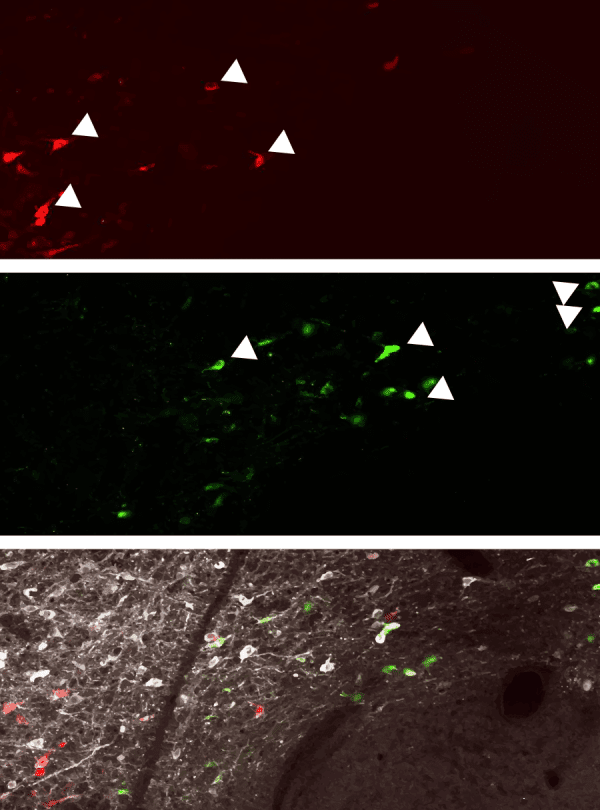
Recent studies have shown that certain groups of neurons in the amygdala are crucial for the regulation of fear responses. The amygdala is a small almond-shaped brain structure in the center of the brain that receives information about fearful stimuli and transmits it to other brain regions to generate fear responses. This causes the body to release stress hormones, change heart rate or trigger fight, flight or freezing responses. Now, a group led by Professors Stéphane Ciocchi of the University of Bern and Andreas Lüthi of the Friedrich Miescher Institute in Basel has discovered that the amygdala plays a much more active role in these processes than previously thought: Not only is the central amygdala a “hub” to generate fear responses, but it contains neuronal microcircuits that regulate the suppression of fear responses. In animal models, it has been shown that inhibition of these microcircuits leads to long-lasting fear behaviour. However, when they are activated, behaviour returns to normal despite previous fear responses. This shows that neurons in the central amygdala are highly adaptive and essential for suppressing fear. These results were published in the journal Nature Communications.
“Disturbed” suppression leads to long-lasting fear
The researchers led by Stéphane Ciocchi and Andreas Lüthi studied the activity of neurons of the central amygdala in mice during the suppression of fear responses. They were able to identify different cell types that influence the animals’ behaviour. For their study, the researchers used several methods, including a technique called optogenetics with which they could precisely shut down – with pulses of light – the activity of an identified neuronal population within the central amygdala that produces a specific enzyme. This impaired the suppression of fear responses, whereupon animals became excessively fearful. “We were surprised how strongly our targeted intervention in specific cell types of the central amygdala affected fear responses,” says Ciocchi, Assistant Professor at the Institute of Physiology, University of Bern. “The optogenetic silencing of these specific neurons completely abolished the suppression of fear and provoked a state of pathological fear.”
Important for developing more effective therapies
In humans, dysfunction of this system, including deficient plasticity in the nerve cells of the central amygdala described here, could contribute to the impaired suppression of fear memories reported in patients with anxiety and trauma-related disorders. A better understanding of these processes will help develop more specific therapies for these disorders. “However, further studies are necessary to investigate whether discoveries obtained in simple animal models can be extrapolated to human anxiety disorders”, Ciocchi adds.
Original Article: How micro-circuits in the brain regulate fear
More from: University of Bern | Friedrich Miescher Institute for Biomedical Research
The Latest Updates from Bing News & Google News
Go deeper with Bing News on:
Suppression of fear responses
- USC decision to deploy police on Gaza war protesters distorts public perception against students
The group’s demands of USC are, in brief: divestment from Israel, cutting off the police presence on campus, ending the university’s displacement of South Central LA residents and denouncing the ...
- Protests continue at Virginia Tech after arrests Sunday night
Dozens of protestors were taken away in handcuffs Sunday night from the student-led Gaza-liberation encampment on Virginia Tech's campus.
- Violent arrest of Emory Professor spotlights brutality of police crackdown on campus protests
The arrest of Emory University economics professor Caroline Fohlin has ignited a firestorm of controversy and drawn widespread attention to what many are calling an excessive use of force by police ...
- PEN America Has Stood By Authors. They Should Stand By PEN.
In response and in keeping with its mission of independence and free expression, PEN America accepted the writers’ willingness to voice their conscience. It has also made clear that there is room for ...
- Local environmentalist joins factory-farm pollution lawsuit against EPA
NORTH CAROLINA — Cape Fear River Watch’s designated Riverkeeper is petitioning the Environmental Protection Agency to enforce protections for an industry he believes is at the heart of local ...
Go deeper with Google Headlines on:
Suppression of fear responses
[google_news title=”” keyword=”suppression of fear responses” num_posts=”5″ blurb_length=”0″ show_thumb=”left”]
Go deeper with Bing News on:
Human anxiety disorders
- Have smartphones created an ‘anxious generation’ among teenagers?
A large majority of Gen Z do not have anxiety disorders – and of those who do ... Haidt explains that, normally, for almost all human history and evolution, these incentives drove teenagers to do ...
- Research consortium to scour the brain for causes of psychiatric illnesses
Science X is a network of high quality websites with most complete and comprehensive daily coverage of the full sweep of science, technology, and medicine news ...
- Sara Evans Talks Struggle with an Eating Disorder, Body Dysmorphia: 'That's Not Normal'
Country singer Sara Evans spoke to Cheryl Burke about struggling with an eating disorder and body dysmorphia, admitting that she’s “scared of being fat” and she knows that’s “not normal” to think.
- How anxiety became a catchall for every unpleasant emotion
When you run a therapy practice called the Center for Anxiety, as David H. Rosmarin does, you encounter a breadth of anxiety-related experiences. Sometimes, after talking with new patients, Rosmarin ...
- The Internet Has Made Health Anxiety Worse Than Ever
I was confused because the opportunities for me to use the internet to research my recent diagnosis of Hodgkin’s Lymphoma, a kind of blood cancer, were minimal anyway. I didn’t own a smartphone or a ...
Go deeper with Google Headlines on:
Human anxiety disorders
[google_news title=”” keyword=”human anxiety disorders” num_posts=”5″ blurb_length=”0″ show_thumb=”left”]
