


CREDIT
Dartmouth College
Dartmouth engineers and physicians develop innovative stereovision system
Researchers at Dartmouth College have found a way to make back surgery safer, faster and more cost effective.
MRIs and CT scans help surgeons identify spine problems, like compressed vertebrae or herniated disks, but finding a clear path to those problem areas is not always as straightforward. Tissue and bone not only stand in the way, they can also move during spinal surgery, rendering a CT scan taken prior to surgery much less accurate.
To solve this problem, Dartmouth professors from the Thayer School of Engineering and the Geisel School of Medicine developed a 3-dimensional, real-time optical tracking system to guide back surgeons as they operate, like a Google Maps for the body, according to findings published in the journal Operative Neurosurgery.
Using a complex software algorithm and two cameras attached to a surgical microscope, the system produces real-time 3-dimensional digitized images on a monitor, according to the study. This type of tracked, calibrated stereoscopic camera system has been extensively used in brain surgery but until now has been unexplored for use in spinal surgery.
The surgeon can use this new intraoperative stereovision system (iSV) without any additional radiation or labor-intensive marking of key areas on the patient’s spine, to match up or co-register with the pre-operative CT scan, as some surgeons do today. This new mapping provides more accurate renderings of where spinal implants or other surgical tools and devices need to go during the procedure, and is expected to save up to 30 minutes, according to one of the study’s authors, Keith D. Paulsen, PhD, Robert A. Pritzker Professor of Biomedical Engineering at Thayer School of Engineering at Dartmouth.
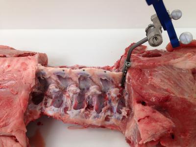
Paulsen and the multidisciplinary team at Dartmouth’s Center for Surgical Innovation tested the new iSV system for accuracy and efficiency while operating on pig spines. Since completing this study, the team has taken its complex system one step further by converting it into a handheld “wand” that the surgeon can pass over the surgical area.
“By rendering images real-time, with a simple handheld tool, we believe we can make surgeries safer and less costly in the future,” said Paulsen.
Next up is fine-tuning the system and testing in humans. The National Institutes of Health has provided the Dartmouth team with another round of funding to continue testing. It could be several years before the system becomes widely available for human spinal surgeries.
Learn more: New imaging system makes back surgery safer, faster and less expensive
The Latest on: Imaging system
[google_news title=”” keyword=”imaging system” num_posts=”10″ blurb_length=”0″ show_thumb=”left”]
via Google News
The Latest on: Imaging system
- New imaging technique provides a much closer look at fibril assemblieson April 26, 2024 at 10:47 pm
A new imaging technique developed by engineers at Washington University in St. Louis can give scientists a much closer look at fibril assemblies, stacks of peptides like amyloid beta, most notably ...
- Medical Digital Imaging Systems Market Elevation Scaling New Heights with Dazzling Trendson April 25, 2024 at 1:50 pm
Check out the latest research report from Report Ocean titled “Medical Digital Imaging Systems Market: Trends Analysis from 2024 to 2032”. This report delivers accurate economic projections, worldwide ...
- Philips receives FDA warning letter over imaging systems manufactured in Chinaon April 25, 2024 at 11:10 am
The FDA published a warning letter that it sent to Philips (NYSE:PHG) that outlines issues around imaging technology manufacturing practices.
- Airborne single-photon lidar system achieves high-resolution 3D imagingon April 25, 2024 at 7:00 am
Researchers have developed a compact and lightweight single-photon airborne lidar system that can acquire high-resolution 3D images with a low-power laser. This advance could make single-photon lidar ...
- Researchers create an AI-powered digital imaging system to speed up cancer biopsy resultson April 25, 2024 at 6:55 am
University of Waterloo researchers have invented a digital medical imaging system that significantly improves the cancer detection process to deliver immediate results and enable swift, effective ...
- Imaging technique shows new details of peptide structureson April 24, 2024 at 11:19 am
A new imaging technique developed by engineers at Washington University in St. Louis can give scientists a much closer look at fibril assemblies—stacks of peptides that include amyloid beta, most ...
- Top 100 U.S. Hospital Health System Selects Intelerad to Deliver Enterprise Imaging Solutionon April 24, 2024 at 6:00 am
Prominent United States health system’s multi-year contract to utilize IntelePACS® with Clario SmartWorklist enables radiology groups to read over two million medical imaging studiesRaleigh, North ...
- Diamond dust shines bright in Magnetic Resonance Imagingon April 23, 2024 at 5:00 pm
An unexpected discovery surprised a scientist: nanometer-sized diamond particles, which were intended for a completely different purpose, shone brightly in a magnetic resonance imaging experiment -- ...
- New imaging system at MUSC provides clear, detailed view for difficult surgerieson April 22, 2024 at 3:59 am
A new digital imaging system at MUSC replaces microscopes for intricate surgeries in the brain and spine. One Summerville family is already thankful for it.
via Bing News

