


Credit: Tanasova, Rao/Michigan Tech
This is not just another tool to image cancer. The probe is a two-for-one: detect cancer and distinguish one type from another. Together, they develop a cancer fingerprint.
Determining the presence of cancer, as well as its type and malignancy, is a stressful process for patients that can take up to two weeks to get a diagnosis. With a new bit of technology—a sugar-transporting biosensor—researchers at Michigan Technological University hope to reduce that timeframe down to minutes.
A collaborative team of chemists and engineers from Michigan Tech lays the groundwork for this vision in two new papers. In the Royal Society of Chemistry’s journal Chemical Communication (DOI: 10.1039/c7cc09809j), the team explains the basic science behind multicolor probes that enable targeting of a cancer-relevant fructose transporter, delving into the image-based detection of cancer cells. In the journal Biosensors (DOI: 10.3390/bios8020039), the team addresses applications for breast cancer detection and differentiating nonmalignant, pre-malignant and malignant cancer cells.
GLUT5 transports fructose and cancer loves sugar
Two Michigan Tech researchers collaborated on the studies. Marina Tanasova, assistant professor of chemistry, and Smitha Rao, assistant professor of biomedical engineering, turned a 10-minute office meeting into a two-year collaboration built on a tiny fluorescent probe that seeks out the fructose transporter named GLUT5.
Cells need carbohydrates; facilitative glucose transporters (GLUTs) bring nutrients in and out of cells. When metabolic swings kick in—say, from cancer development—the overall make-up of GLUTs change so that more or less GLUTs are active. Fructose transporters like GLUT5 are of particular interest because of the direct connection between fructose uptake and cancer development, which also changes as cancer progresses and becomes malignant.
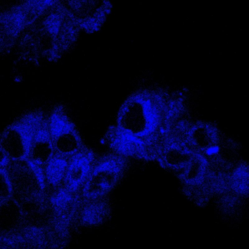
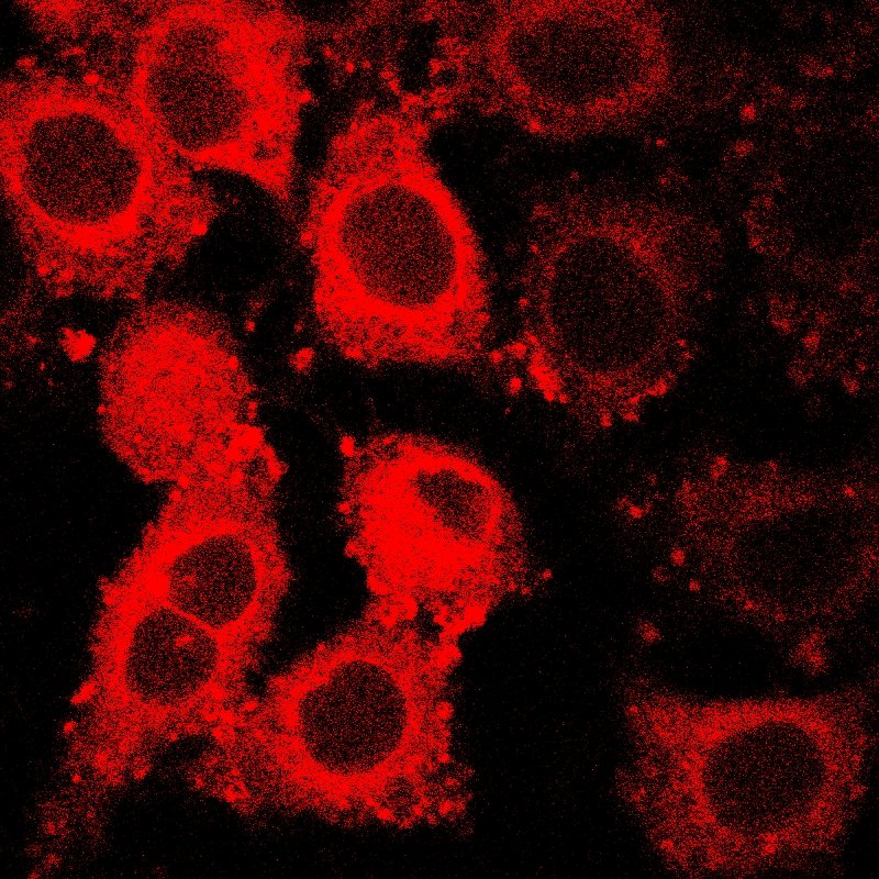
In their Chemical Communications paper, Tanasova and Rao’s team report on the design and validation of the fluorescent probes, called ManCous, to observe GLUT5 activity in a cell. They document the multicolored fluorescence that distinguishes GLUT5-rich cells from those deficient in GLUT5. The follow up Biosensors paper provides a proof-of-principle use of these probes as tools to assess and compare GLUT5 and metabolic activity of different cell types, including normal cells and different cancer subtypes.
“We came closer to a basic screening of cells’ GLUT composition—one that both detects cancer and distinguishes type,” Tanasova says, adding that while the concept is elegant, developing the technology is not easy. “From basic science to application, there is a lot of transformation that has to happen.”
Engineering better cancer detection takes collaboration
By better understanding the science behind sugar transporters, the team is more equipped to build technology that captures an accurate and precise GLUT fingerprint of cancerous cells.
“This probe is like a Swiss army knife,” Rao says, explaining that cancer detection is not the only use for the probe. “The more we learn about cancer through these probes, the more opportunities we have to apply them—which means more chances to treat different cancers, hopefully cure them, and at least prevent their spread and maximize drug delivery.”

What makes the probe so versatile is its ability to not only seek out and highlight cancer cells, but also to reveal the metabolic nuances of different stages of cancer development. While cancer generally gobbles up fructose, the GLUT5 and metabolic activity of nonmalignant, pre-malignant and malignant breast cancer cells do vary, which is what Rao, Tanasova and their team explored in the Biosensors paper. They found that ManCou probes allow for parallel analysis and quantification of GLUT5 and metabolic activity of cells just after a 10-minute incubation period. The notable differences in fluorescence intensity observed between normal cells and cancer cells, as well as different cancer types, provide important points of differentiation between different cell types and make these probes promising tools for cancer detection and diagnostics.
Learn more: GLUT5 Fluorescent Probe Fingerprints Cancer Cells

The Latest on: Cancer fingerprint
[google_news title=”” keyword=”cancer fingerprint” num_posts=”10″ blurb_length=”0″ show_thumb=”left”]
via Google News
The Latest on: Cancer fingerprint
- Unique fingerprint of cancer cells revealed in hydrogen atomson May 7, 2024 at 6:00 am
Geologists and biologists use earth science tools to identify cancer's unique atomic markers known as fingerprints.
- Scientists Reveal Cancer's Atomic Secrets—'Whole New Layer to Medicine'on May 7, 2024 at 5:44 am
By studying these ratios in yeast and mouse liver cells, the team found that cells that were growing extremely fast, similar to cancer cells, contained a noticeably higher ratio of hydrogen to ...
- Geological tool helps study biology of cancer at atomic levelson May 6, 2024 at 12:00 pm
A groundbreaking study reveals cancer cells have a distinct hydrogen composition, potentially revolutionizing early detection.
- Geologists, biologists unearth the atomic fingerprints of canceron May 5, 2024 at 5:00 pm
Scientists at the University of Colorado Boulder and Princeton University have, for the first time, employed a tool often used in geology to detect the atomic fingerprints of cancer. In a case of ...
- Pancreatic Cancer Is Hard to Detect in Early Stages. A New Blood Test Could Helpon May 3, 2024 at 11:46 am
A liquid biopsy can detect 97% of stage 1 and 2 pancreatic cancer cases, according to an early clinical trial. Blood tests could help improve early detection for the deadly cancer.
- Unknown risk factor linked to high rates of kidney cancer, DNA study findson May 2, 2024 at 7:22 am
Researchers analyzing the DNA of people with kidney cancer worldwide have found evidence of an unknown trigger that could explain the longstanding mystery of why some countries have a higher incidence ...
- Embracing a New Way of Life After Canceron May 1, 2024 at 5:00 pm
It’s transformed my life. I have discovered my words can help others with their cancer survivorship. All of your hopes, dreams and experiences are like fingerprints in a maze of life. No one has the ...
- New cancer vaccine: Doctor explains all you need to knowon April 26, 2024 at 6:39 am
A doctor has explained more about the world's first personalised mRNA cancer jab for melanoma, which is currently being tested in British patients. Doctor Amir Khan described the vaccine as “positive ...
via Bing News
