


CREDIT
Isabelle Fournier
Isabelle Fournier and her team are out to change surgical oncology.
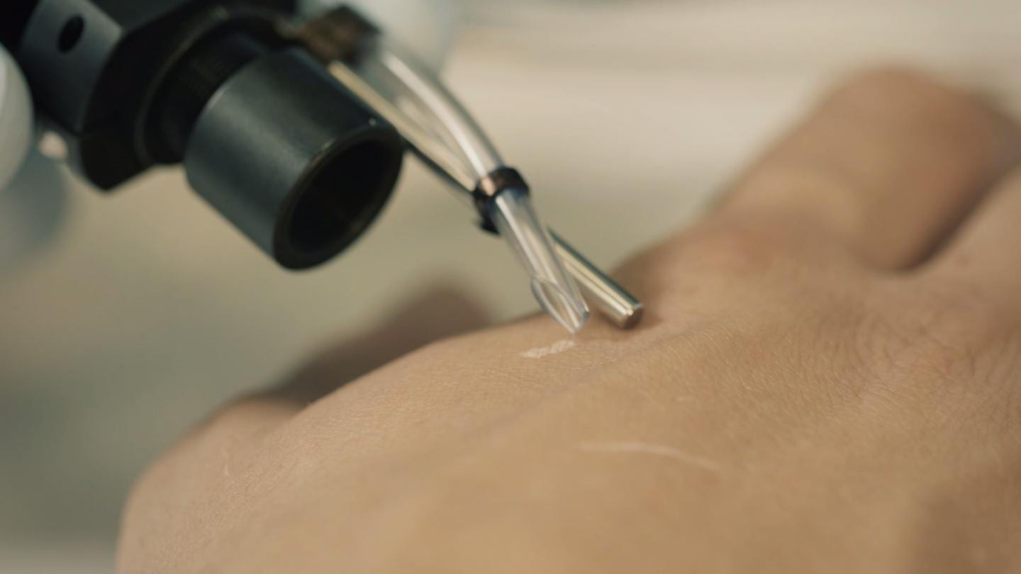
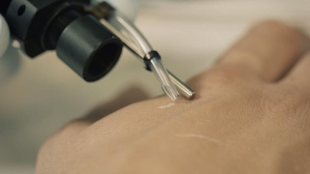
“Better surgery is associated with better prognosis and higher survival,” said Fournier, a professor at the University of Lille and co-director of a proteomics center of INSERM, the French national institute of health. Her laboratory has worked for several years on a device they call the SpiderMass that will enable surgeons to look for markers of cancer in a living patient’s tissue, during an operation. In an article in Molecular & Cellular Proteomics, the team reports on an important step toward finding protein biomarkers during surgery.
Surgery to remove a primary tumor involves a wait. After the tumor and some healthy surrounding tissue are removed, the surgical team pauses while a pathologist checks the tissue margins under a microscope. Although this process is important for preventing recurrence of the cancer, it can add up to 45 risky minutes under anesthesia.
With the new device, Fournier said, “We think that it is possible to open the way to in vivo real-time proteomics,” which could help surgeons find stray cancer cells faster, perhaps even as they make incisions.
Fournier’s device uses mass spectrometry, which measures the mass of molecules from complex mixtures. But turning an in vivo tissue sample into gas phase ions for measurements can be a challenge. Until now, no one knew how to extract ions from living tissues without doing harm.
So Fournier’s team got creative. Riffing on MALDI, an ionization strategy that uses a carrier molecule mixed with the analyte of interest, they decided to use the water that makes up a majority of human tissue as a carrier to produce a water-assisted laser desorption/ionization, or WALDI. If they could excite the water in a tiny area, it should vaporize, taking ionized organic molecules with it.
“It was an idea at the beginning, and many people thought that it would not work,” Fourier said. “Finally, we have it working beautifully.”
The team built a pulsed laser excitation device tuned to heat water precisely by causing vibration in the oxygen-hydrogen bond. In a 2016 paper, they described using this laser to ionize the outermost layer of tissue, penetrating less than one-twentieth of a millimeter. The human volunteers reported a slight tingling sensation. But the ions that appeared were mostly small molecules and lipids, which are more apt than proteins to adopt a negative charge. The team hoped to measure proteins as well.
In this new paper, Fournier and colleagues report that they have cracked the protein puzzle. By using a more sensitive mass spectrometer and looking for positively instead of negatively charged ions, they found peaks representing purified proteins they had introduced into a cow liver sample. Now that they know the proteins are detectable, the next step will be finding ways to amplify the protein signal over more abundant lipids and metabolites.
In the meantime, the device is already in use for four-legged patients. Fournier’s lab has worked with the veterinary biotech company Oncovet Clinical Research to run a pilot trial, comparing biopsies from pet dogs with sarcoma to healthy tissues. The team developed a lipidomics- and metabolomics-based classification system to robustly identify healthy, necrotic and cancerous tissues. Soon, they will introduce a prototype into a veterinary operating room. If it is successful there, Fournier said, she hopes to reach human clinics, improving tumor removal surgery to give patients better health outcomes.
Learn more: Painless real-time proteomics may one day speed up cancer surgery

The Latest on: Proteomics
[google_news title=”” keyword=”proteomics ” num_posts=”10″ blurb_length=”0″ show_thumb=”left”]
via Google News
The Latest on: Proteomics
- New multi-task deep learning framework integrates large-scale single-cell proteomics and transcriptomics dataon April 26, 2024 at 7:35 am
The exponential progress in single-cell multi-omics technologies has led to the accumulation of large and diverse multi-omics datasets. However, the integration of single-cell proteomics and ...
- Acrivon Therapeutics’ shares climb 14% on plans to present cancer-test dataon April 24, 2024 at 4:16 pm
Shares of Acrivon Therapeutics advanced in post-market trading after the biopharmaceutical company said it would present clinical trial data in connection with a biomarker test for ovarian and ...
- Acrivon Therapeutics Reports Initial Positive Clinical Data for ACR-368 and Pipeline Program Progress Today at Corporate R&D Eventon April 24, 2024 at 1:25 pm
Initial ACR-368 Phase 2b clinical data in patients with ovarian or endometrial cancers (n=26; 10 OncoSignature-positive and 16 ...
- Identifying proteins causally related to COVID-19, healthspan and lifespanon April 24, 2024 at 1:05 pm
A new research paper titled "Using genetics and proteomics data to identify proteins causally related to COVID-19, healthspan and lifespan: a Mendelian randomization study" has been published in Aging ...
- Proteomics News and Researchon April 22, 2024 at 4:59 pm
A new case report published in the peer-reviewed OMICS: A Journal of Integrative Biology describes how longitudinal multi-omics monitoring (LMOM) helped to detect a precancerous pancreatic tumor ...
- Proteomics Moves From Expression to Turnoveron April 22, 2024 at 4:59 pm
Measuring protein turnover is complex. Even in simple unicellular organisms such as bacteria and yeast, there are many up- and down-stream processes that must be considered. The major premise of ...
- Visual Proteomics: Observing Protein Location in Living Cellson April 19, 2024 at 6:00 am
Proteins function dynamically in cells, and tracking their location precisely in living cells has been a major technical challenge. Current methods involve individually labeling individual proteins ...
- Proteomics Market Expanding Rapidly, Predicted Value of USD 103.8 Billion by 2032on April 14, 2024 at 5:00 pm
According to a recent report by Market.us, the Global Proteomics Market size is expected to be worth around USD 103.8 Billion by 2032 from USD 30.8 Billion in 2023, growing at a CAGR of 14.9% during ...
- Structural proteomics of an archaeonon March 19, 2024 at 8:33 am
The completion and near completion of the sequencing phase of genome projects has ushered in the age of proteomics, the study of all gene products in an organism. This flood of sequence ...
via Bing News
