


(Credit: Ian Papautsky)
Researchers at the University of Illinois at Chicago and Queensland University of Technology of Australia, have developed a device that can isolate individual cancer cells from patient blood samples.
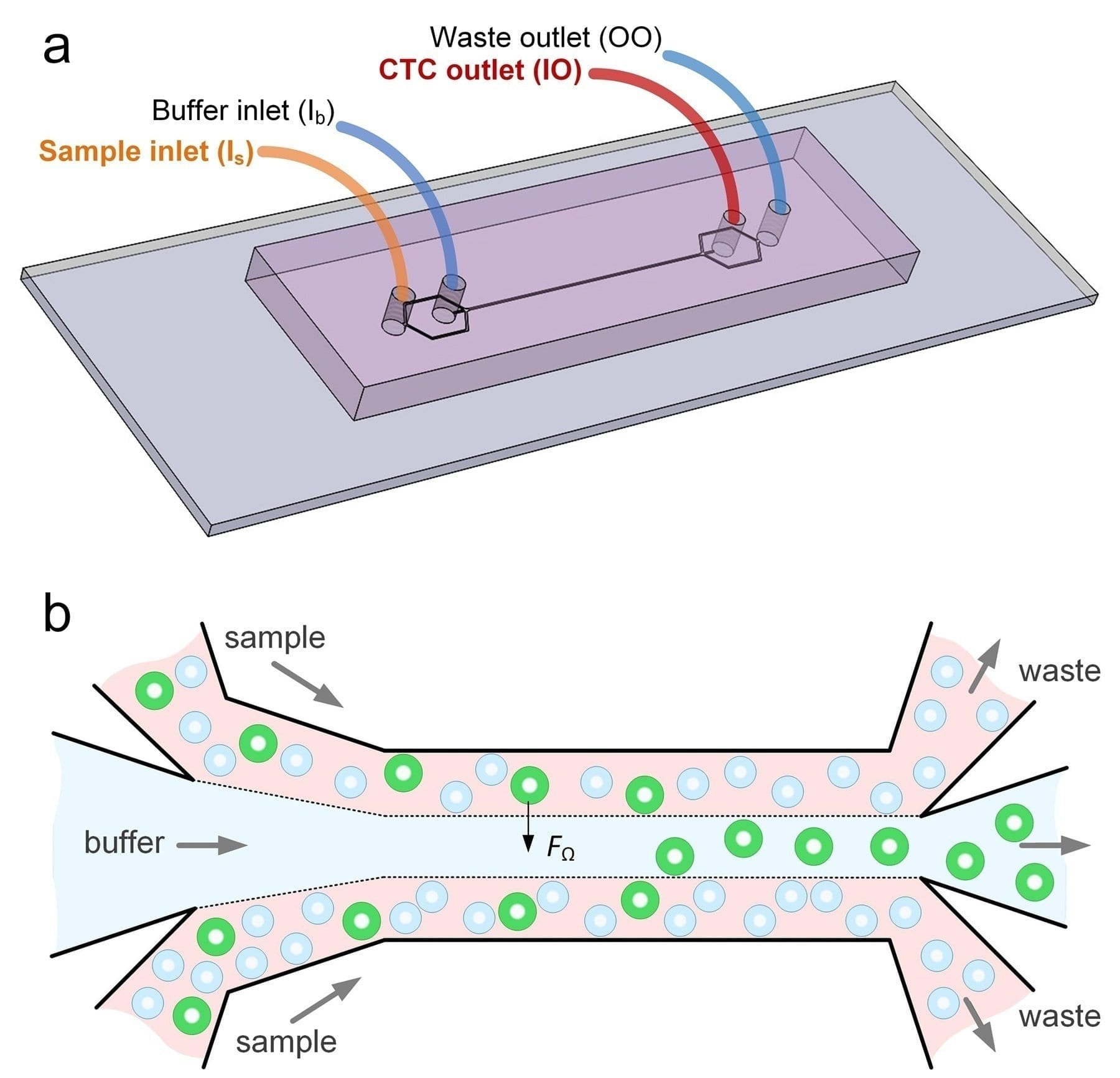
The microfluidic device works by separating the various cell types found in blood by their size. The device may one day enable rapid, cheap liquid biopsies to help detect cancer and develop targeted treatment plans. The findings are reported in the journal Microsystems & Nanoengineering.
“This new microfluidics chip lets us separate cancer cells from whole blood or minimally-diluted blood,” said Ian Papautsky, the Richard and Loan Hill Professor of Bioengineering in the UIC College of Engineering and corresponding author on the paper. “While devices for detecting cancer cells circulating in the blood are becoming available, most are relatively expensive and are out of reach of many research labs or hospitals. Our device is cheap, and doesn’t require much specimen preparation or dilution, making it fast and easy to use.”
The ability to successfully isolate cancer cells is a crucial step in enabling liquid biopsy where cancer could be detected through a simple blood draw. This would eliminate the discomfort and cost of tissue biopsies which use needles or surgical procedures as part of cancer diagnosis. Liquid biopsy could also be useful in tracking the efficacy of chemotherapy over the course of time, and for detecting cancer in organs difficult to access through traditional biopsy techniques, including the brain and lungs.
However, isolating circulating tumor cells from the blood is no easy task, since they are present in extremely small quantities. For many cancers, circulating cells are present at levels close to one per 1 billion blood cells. “A 7.5-milliliter tube of blood, which is a typical volume for a blood draw, might have ten cancer cells and 35-40 billion blood cells,” said Papautsky. “So we are really looking for a needle in a haystack.”
Microfluidic technologies present an alternative to traditional methods of cell detection in fluids. These devices either use markers to capture targeted cells as they float by, or they take advantage of the physical properties of targeted cells — mainly size — to separate them from other cells present in fluids.
Papautsky and his colleagues developed a device that uses size to separate tumor cells from blood. “Using size differences to separate cell types within a fluid is much easier than affinity separation which uses ‘sticky’ tags that capture the right cell type as it goes by,” said Papautsky. “Affinity separation also requires a lot of advanced purification work which size separation techniques don’t need.”
Learn more: New microfluidics device can detect cancer cells in blood
The Latest on: Cancer blood test
[google_news title=”” keyword=”cancer blood test” num_posts=”10″ blurb_length=”0″ show_thumb=”left”]
via Google News
The Latest on: Cancer blood test
- CU Cancer Center Helps Lead National Trial to Evaluate Multicancer Blood Testson April 26, 2024 at 9:38 am
The Vanguard Study will evaluate whether the benefits of using blood tests to screen for multiple cancers outweigh the harms.
- Bay Area tech layoffs: Cancer screening company to cut 20% of workforce after securing $254 millionon April 24, 2024 at 4:44 pm
Freenome, a South San Francisco biotechnology company, plans to reduce its workforce by approximately 20%. The decision will impact over 100 employees across various departments, part of an effort to ...
- Company developing early cancer-detection tests to cut 20% of workforceon April 24, 2024 at 7:35 am
Early cancer-detection test maker that raised $254 million earlier this year now says it will cut more than 100 jobs — about 20% of its workforce — as it restructures ...
- New Test Detects Cancer In Minutes Using Just 1 Drop of Dried Bloodon April 24, 2024 at 6:04 am
A test devised in Shanghai could make cancer detection quicker, easier, and practical for multiple areas of the body at once. Using just one drop of dried blood, the test looks for biomarkers that ...
- Balancing hope and reality: The promise and peril of blood-based colorectal cancer screeningon April 24, 2024 at 1:30 am
A simple blood test to detect colorectal cancer sounds amazing. But unlike colonoscopy, a blood test can't remove precancerous polyps.
- AI-powered tool detects cancer in minutes with one drop of bloodon April 23, 2024 at 6:52 am
Scientists in China have developed a test that detects three types of cancers with just one spot of dried blood.
- Researchers developing new colon cancer tests amid rising caseson April 23, 2024 at 3:01 am
With more than 200,000 cases of colon cancer diagnosed across the country every year, researchers are working on new ways to detect the disease better.
- Detecting cancer in minutes possible with just a drop of dried blood and new test, study hintson April 22, 2024 at 9:19 am
Early tests suggest that a new tool that requires only a single drop of blood could detect three of the deadliest forms of cancer.
- New Urine Test for Prostate Cancer May Help Reduce Unnecessary Biopsieson April 22, 2024 at 4:19 am
A prostate cancer biomarker test that utilizes genes indicating high-grade prostate cancer appears highly accurate.
- Simple urine test ‘could help avoid unnecessary prostate cancer biopsies’on April 18, 2024 at 4:00 am
At present, tests to check for prostate cancer include a digital rectal examination, a PSA blood test, MRI scans and a sample, or biopsy, of the prostate gland. But blood tests or MRI alone cannot ...
via Bing News
