


 UBC researchers develop new laser microscope that could be ‘revolutionary’ for treatment of diseases such as skin cancer
University of British Columbia researchers have developed a specialized microscope that has the potential ability to both diagnose diseases that include skin cancer and perform incredibly precise surgery—all without cutting skin.
The researchers describe the technology in a study published today in Science Advances.
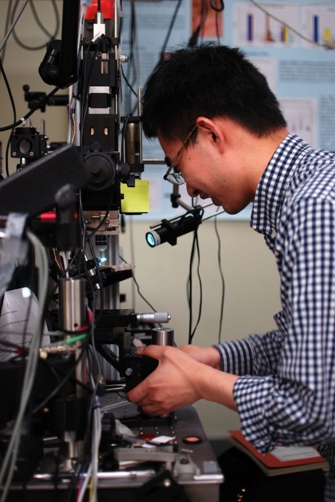
“Our technology allows us to scan tissue quickly, and when we see a suspicious or abnormal cell structure, we can perform ultra-precise surgery and selectively treat the unwanted or diseased structure within the tissue—without cutting into the skin,” said Yimei Huang, co-lead author of the study and a former postdoctoral fellow at the department of dermatology and skin science at UBC and BC Cancer.
Huang co-led the study with Zhenguo Wu, a UBC PhD student.
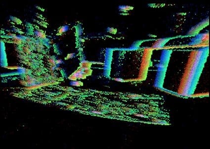
The device is a specialized type of multiphoton excitation microscope that allows imaging of living tissue up to about one millimeter in depth using an ultrafast infrared laser beam. What sets the researchers’ microscope apart from previous technology is that it’s capable of not only digitally scanning living tissue, but also treating the tissue by intensifying the heat produced by the laser.
When applied to treating diseases of the skin, the microscope allows medical professionals to pinpoint the exact location of the abnormality, diagnose it and treat it instantly. It could be used to treat any structure of the body that is reached by light and that requires extremely precise treatment, including nerves or blood vessels in the skin, eye, brain or other vital structures.
“We can alter the pathway of blood vessels without impacting any of the surrounding vessels or tissues,” said study co-author Harvey Lui, professor at the department of dermatology and skin science at UBC and the Vancouver Coastal Health Research Institute, and a dermatologist at BC Cancer. “For diagnosing and scanning diseases like skin cancer, this could be revolutionary.”
The researchers wanted to make multiphoton microscope technology more versatile while also increasing its precision.
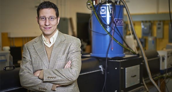
“We wanted to be able to identify what was happening under the skin from many different angles and to have the capability of imaging different body sites,” said senior author Haishan Zeng, professor of dermatology, pathology and physics at UBC and distinguished scientist with BC Cancer.
“Once we achieved that, we wondered whether we could transform this diagnostic device into a treatment device by simply turning up the power of the laser.”
The results were incredibly exciting.
“We are not only the first to achieve fast video-rate imaging that enables clinical applications, but also the first to develop this technology for therapeutic uses,” said Zeng.
The researchers have partnered with several UBC departments, including mechanical engineering, electrical engineering and ophthalmology, to develop different versions of the technology. Exploration includes research into the development of a miniature version that could be used to perform microscopic examinations and treatment during endoscopy—a non-surgical procedure used to examine a person’s digestive tract using an endoscope, a flexible tube with a light and camera attached to it.
Learn more: A new way of diagnosing and treating disease—without cutting skin
The Latest on: Laser microscope
[google_news title=”” keyword=”laser microscope” num_posts=”10″ blurb_length=”0″ show_thumb=”left”]
via Google News
The Latest on: Laser microscope
- Challenging Old Theories: Innovative Microscopy Exposes New Alzheimer’s Treatment Pathwayson April 27, 2024 at 3:35 am
Researchers at UC San Diego have utilized advanced imaging techniques to explore the metabolic processes behind Alzheimer's disease, leading to potential new strategies for treatment. Alzheimer's ...
- Exploring gel formation mechanisms and the role of lactic acid bacteria in fermented sausageon April 25, 2024 at 10:22 am
A research team has reviewed the process of gel formation in fermented sausages, emphasizing the crucial role of myofibrillar proteins and the influence of lactic acid bacteria, temperature, and ...
- Scientists pioneer new X-ray microscopy method for data analysis 'on the fly'on April 24, 2024 at 9:54 am
A new streaming technique allows playback of data while it is being generated. When scientists want to look at a tiny structure in a material, even one just a few atoms in size, they frequently turn ...
- ICFO develops QUIONE, quantum simulator that observes individual atoms…on April 24, 2024 at 4:37 am
…and Toshiba-Single Quantum collaboration doubles range of secure QKD communications.
- Innovative microscopy demystifies metabolism of Alzheimer'son April 23, 2024 at 3:56 pm
Using state-of-the-art microscopy techniques, researchers have shed new light on the underlying mechanisms driving Alzheimer's disease.
- World’s only quantum-gas microscope imaging strontium’s individual atomson April 23, 2024 at 4:40 am
Spanish researchers develop the world's first quantum-gas microscope that captures images of individual atoms of strontium quantum gases.
- SkyWater Technology Is Lowering Barriers To Advanced Chip Packagingon April 23, 2024 at 3:00 am
SkyWater has partnered with Deca Technologies to develop a multi chip packaging capability that will lower the entry barriers to advanced packaging.
- Announcing the birth of QUIONE, a unique analog quantum processoron April 22, 2024 at 1:31 pm
Quantum physics requires high-precision sensing techniques to delve deeper into the microscopic properties of materials. From the analog quantum processors that have emerged recently, quantum-gas ...
- Hybrid camera system gives your smartphone "super-telephoto" zoomon April 18, 2024 at 5:43 am
Tech company Beaverlab has launched a Kickstarter to raise funds for a smart super-telephoto camera called the Excope DT1 that can be had with a 400-mm zoom lens, and works with a smartphone to ...
- A better view with new mid-infrared nanoscopyon April 17, 2024 at 2:42 am
A team at the University of Tokyo have constructed an improved mid-infrared microscope, enabling them to see the structures inside living bacteria at the nanometer scale. Mid-infrared microscopy is ...
via Bing News
