


Microscopy system is faster, simpler, and cheaper
Opening new doors for biomedical and neuroscience research, Elizabeth Hillman, associate professor of biomedical engineering at Columbia Engineering and of radiology at Columbia University Medical Center (CUMC), has developed a new microscope that can image living things in 3D at very high speeds. In doing so, she has overcome some of the major hurdles faced by existing technologies, delivering 10 to 100 times faster 3D imaging speeds than laser scanning confocal, two-photon, and light-sheet microscopy. Hillman’s new approach uses a simple, single-objective imaging geometry that requires no sample mounting or translation, making it possible to image freely moving living samples. She calls the technique SCAPE, for swept confocally aligned planar excitation microscopy. Her study is published in the Advance Online Publication (AOP) on Nature Photonics’s website on January 19, 2015.
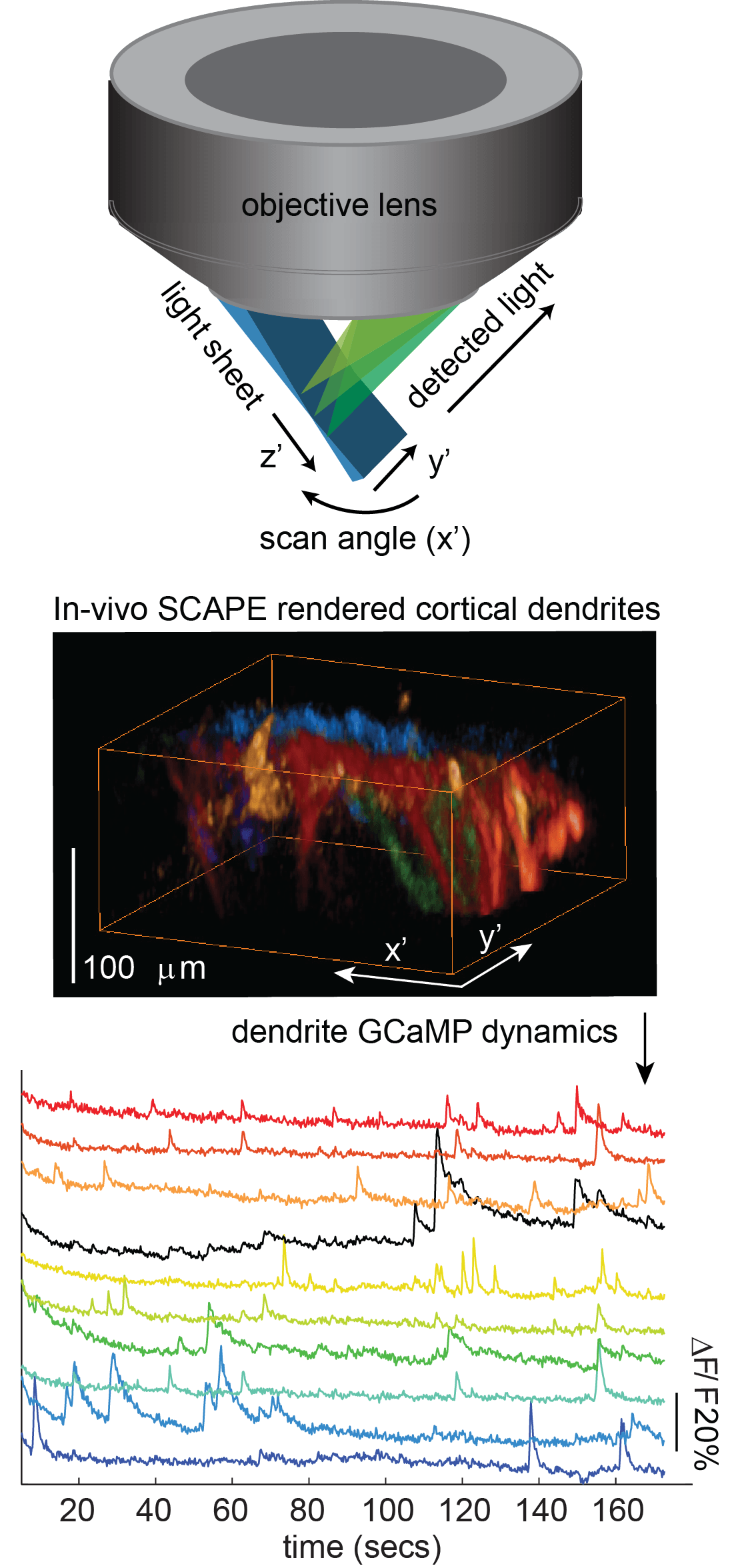
“The ability to perform real-time 3D imaging at cellular resolution in behaving organisms is a new frontier for biomedical and neuroscience research,” says Hillman, who is also a member of Columbia’s Mortimer B. Zuckerman Mind Brain Behavior Institute. “With SCAPE, we can now image complex, living things, such as neurons firing in the rodent brain, crawling fruit fly larvae, and single cells in the zebrafish heart while the heart is actually beating spontaneously—this has not been possible until now.”
Highly aligned with the goals of President Obama’s BRAIN Initiative, SCAPE is a variation on light-sheet imaging, but, “It breaks all the rules,” says Hillman. While conventional light-sheet microscopes use two awkwardly positioned objective lenses, Hillman realized that she could use a single-objective lens, and then that she could sweep the light sheet to generate 3D images without moving the objective or the sample. “This combination makes SCAPE both fast and very simple to use, as well as surprisingly inexpensive,” she explains. “We think it will be transformative in bringing the ability to capture high-speed 3D cellular activity to a wide range of living samples.”
SCAPE is an urgently needed breakthrough.
Read more: New High-Speed 3D Microscope—Scape—Gives Deeper View of Living Things
The Latest on: 3D Microscope
[google_news title=”” keyword=”3D Microscope” num_posts=”10″ blurb_length=”0″ show_thumb=”left”]
via Google News
The Latest on: 3D Microscope
- Scientists create 3D reconstruction of a millimetre-sized fragment of human brain tissueon May 10, 2024 at 3:05 am
They used electron microscope images and artificial intelligence to colour-code and reconstruct the brain tissue in 3D. Their reconstruction contains roughly 57,000 cells, about 230 millimetres of ...
- MIT Technology Reviewon May 9, 2024 at 4:20 pm
A small brain sample was sliced into 5,000 pieces, and machine learning helped stitch it back together.
- BIZTODAY China-Super Microscopeon May 2, 2024 at 8:54 pm
Chinese researchers unveil new generation of super microscope 【Voice_over】 A group of researchers from Tsinghua University in Beijing recently unveiled a next generation light field microscope. These ...
- New technology makes 3D microscopes easier to use, less expensive to manufactureon April 30, 2024 at 8:45 am
D microscopes are used in applications from the life sciences to semiconductor manufacturing. Now engineers are developing patented and patent-pending innovations making them work faster to capture ...
- Purdue University 3D microscope innovations (IMAGE)on April 30, 2024 at 7:43 am
Liming Chen, who will earn his PhD in mechanical engineering from Purdue in May 2024, operates a 3D microscope, which uses an optical technique called fringe projection to create a high-resolution 3D ...
- Purdue-created technology makes 3D microscopes easier to use, less expensive to manufactureon April 30, 2024 at 7:26 am
Liming Chen, who will earn his PhD in mechanical engineering from Purdue in May 2024, operates a 3D microscope, which uses an optical technique called fringe projection to create a high-resolution ...
- First high-resolution 3D nanoscale chemical imaging achieved with multi-modal tomographyon April 30, 2024 at 6:21 am
By exploiting a smart learning algorithm that fuses two microscopy signals, University of Michigan researchers have accomplished high-resolution, efficient 3D chemical imaging for the first time at ...
- Image Shows What Caffeine Looks Like Under Microscope?on April 29, 2024 at 4:00 am
"That's what I see when I get a migraine from lack of or drinking too much caffeine," one Reddit user commented.
- ZEISS Xradia 410 Versa 3D X-Ray Microscope from Carl Zeisson April 26, 2024 at 5:00 pm
The ZEISS Xradia 410 Versa encompasses all the benefits of the VersaXRM family of 3D X-ray microscopes for computed tomography. Xradia 410 Versa is uniquely designed to bridge cost/performance ...
- NPFLEX 3D Optical Microscope from Brukeron April 26, 2024 at 5:00 pm
The NPFLEX™ provides the most flexible, non-contact, 3D areal surface characterization for such large samples as orthopedic medical implants and the larger parts in aerospace, automotive and ...
via Bing News

