


Researchers at The University of Nottingham have developed a break-through technique that uses sound rather than light to see inside live cells, with potential application in stem-cell transplants and cancer diagnosis.
The new nanoscale ultrasound technique uses shorter-than-optical wavelengths of sound and could even rival the optical super-resolution techniques which won the 2014 Nobel Prize for Chemistry.
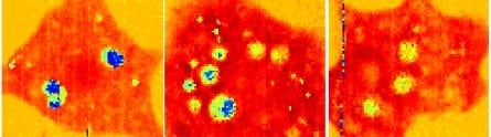
This new kind of sub-optical phonon (sound) imaging provides invaluable information about the structure, mechanical properties and behaviour of individual living cells at a scale not achieved before.
Researchers from the Optics and Photonics group in the Faculty of Engineering, University of Nottingham, are behind the discovery, which is published in the paper ‘High resolution 3D imaging of living cells with sub-optical wavelength phonons’ in the journal, Scientific Reports.
“People are most familiar with ultrasound as a way of looking inside the body — in the simplest terms we’ve engineered it to the point where it can look inside an individual cell. Nottingham is currently the only place in the world with this capability,” said Professor Matt Clark, who contributed to the study.
In conventional optical microscopy, which uses light (photons), the size of the smallest object you can see (or the resolution) is limited by the wavelength.
For biological specimens, the wavelength cannot go smaller than that of blue light because the energy carried on photons of light in the ultraviolet (and shorter wavelengths) is so high it can destroy the bonds that hold biological molecules together damaging the cells.
Optical super-resolution imaging also has distinct limitations in biological studies. This is because the fluorescent dyes it uses are often toxic and it requires huge amounts of light and time to observe and reconstruct an image which is damaging to cells.
Unlike light, sound does not have a high-energy payload. This has enabled the Nottingham researchers to use smaller wavelengths and see smaller things and get to higher resolutions without damaging the cell biology.
“A great thing is that, like ultrasound on the body, ultrasound in the cells causes no damage and requires no toxic chemicals to work. Because of this we can see inside cells that one day might be put back into the body, for instance as stem-cell transplants,” adds Professor Clark.
Learn more: New ultrasound technique is first to image inside live cells
[osd_subscribe categories=’ultrasound’ placeholder=’Email Address’ button_text=’Subscribe Now for any new posts on the topic “ULTRASOUND”‘]
Receive an email update when we add a new ULTRASOUND article.
The Latest on: Nanoscale ultrasound
[google_news title=”” keyword=”nanoscale ultrasound” num_posts=”10″ blurb_length=”0″ show_thumb=”left”]
via Google News
The Latest on: Nanoscale ultrasound
- Diamond dust shines bright: A safer contrast agent for MRI scanson April 27, 2024 at 1:54 am
Nanoscale diamonds unexpectedly shine brighter in MRI scans, sparking research into their potential as a novel contrast agent.
- New imaging technique provides a much closer look at fibril assemblieson April 26, 2024 at 10:47 pm
A new imaging technique developed by engineers at Washington University in St. Louis can give scientists a much closer look at fibril assemblies, stacks of peptides like amyloid beta, most notably ...
- Sound and vision: synchrotron insights illuminate crystal nucleation and growthon April 24, 2024 at 7:44 am
Curiosity-driven research using low-power ultrasound fields to investigate the fundamental physics of crystal nucleation – the formation of crystal nuclei and “embryos” in the liquid or solution phase ...
- Imaging technique shows new details of peptide structureson April 23, 2024 at 5:00 pm
A new imaging technique developed by engineers at ... "We engineer microscopes to enable better nanoscale measurements so that the science can move forward," Lew said. In a paper published ...
- Realizing the Biological and Biomedical Potential of Nanoscale Imaging Using a Pipette Probeon April 18, 2024 at 5:00 pm
Ion conductance microscopy can scan complex cellular structures and tissues with nanoscale resolution. A suite of different imaging modalities can be performed with the same instrument in order to ...
- Atom-by-atom: Imaging structural transformations in 2D materialson April 17, 2024 at 2:53 pm
Silicon-based electronics are approaching their physical limitations and new materials are needed to keep up with current technological demands. Two-dimensional (2D) materials have a rich array of ...
- STED Microscopy: Revolutionizing Nanoscale Imaging in Biology and Beyondon April 15, 2024 at 1:09 pm
Stimulated Emission Depletion (STED) microscopy is a super-resolution imaging technique that overcomes the diffraction limit of conventional optical microscopy. It enables the visualization of ...
- Nanoscale movies shed light on one barrier to a clean energy futureon April 10, 2024 at 5:00 pm
Nanoscale movies shed light on one barrier to a clean energy future. ScienceDaily . Retrieved April 22, 2024 from www.sciencedaily.com / releases / 2024 / 04 / 240411130341.htm ...
- Nanotechnology Newson April 10, 2024 at 5:00 pm
Using nanoscale imaging techniques, researchers have captured high-resolution videos of tiny crystals of ruthenium ...
via Bing News

