


Research quality X-rays could have widespread applications
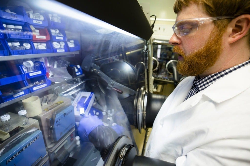
Using a compact but powerful laser, a research team at the University of Nebraska-Lincoln has developed a new way to generate synchrotron X-rays.
Although the high quality of synchrotron X-rays make them ideal for research ranging from the structure of matter to advanced medical images, access to the technology has been limited until now. Most traditional synchrotron X-ray devices are gigantic and costly, available only at a few sites around the world.
As reported in this week’s issue of the top-ranked optics journal Nature Photonics, researchers at UNL’s Extreme Light Laboratory developed a novel method to generate research-quality X-rays using a “tabletop” laser.
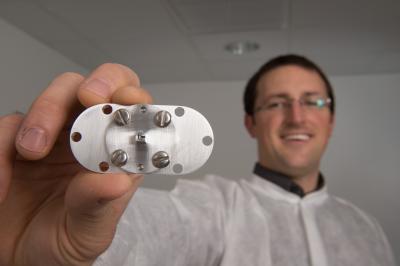
“Our hope is that this new technology will lead to applications that benefit both science and society,” said Nathan Powers, a Ph.D. student and first author of the journal article.
Physics professor Donald Umstadter, director of the Extreme Light Laboratory, led the research project. He compared the synchrotron X-ray breakthrough to the development of personal computers, giving more people access to computing power once available only via large and costly mainframe computers. Shrinking components of advanced laser-based technology will increase the feasibility of producing high-quality X-rays in medical and university research laboratories, which in turn could lead to new applications for the X-rays.
Because the new X-ray device could be small enough to fit in a hospital or on a truck, it could lead to more widespread applications for advanced X-ray technology, UNL scientists said. New applications might include Homeland Security detecting nuclear materials concealed within a shielded container; doctors finding cancerous tumors at earlier stages; or scientists studying extremely fast reactions that occur too rapidly for observation with conventional X-rays.
Ever since synchrotron X-ray light sources were developed more than 60 years ago, they have grown in size. Some now equal the size of a college campus, with a cost in the hundreds of millions of dollars. These huge machines continue to be built, most recently in Australia and Brazil.
Like supercomputers, they provide scientists with the most advanced research capabilities, yet they are not feasible for most practical applications. Though synchrotron X-rays result in lower doses of radiation as well as high-quality images, the tens of thousands of compact X-ray devices currently in operation in hospitals or at ports worldwide produce lower quality X-rays.
The Latest Bing News on:
X-ray device
- New security cameras, mail screening devices: State to spend millions on security at correctional facilitieson April 27, 2024 at 8:04 pm
The state is looking at spending millions of dollars to beef up security at Hawaii's prisons. This comes after reports of sexual abuse in the past and smuggling of illegal drugs into facilities.
- Revolutionizing Photography with Robopic: The Future of AI-Powered Imagingon April 27, 2024 at 8:33 am
In a world where visual content reigns supreme, the demand for high-quality, captivating imagery has never been ...
- New X-ray machine officially opens at hospitalon April 27, 2024 at 12:58 am
A new X-ray machine room has been officially opened following a ten year fundraising campaign. The modern facility at Aldeburgh Community Hospital, in Suffolk, was built using a gift of £320,000 from ...
- Philips receives FDA warning letter over imaging systems manufactured in Chinaon April 25, 2024 at 11:10 am
The FDA published a warning letter that it sent to Philips (NYSE:PHG) that outlines issues around imaging technology manufacturing practices.
- Medical Imaging Devices Market: Today and Over the Next 10 Years (2024-2034)on April 25, 2024 at 6:47 am
The "Medical Imaging Devices Market Report 2024-2034" report has been added to ResearchAndMarkets.com's offering.Overall world revenue for Medical Imaging Devices Market, 2023 to 2034 in terms of ...
- Trailblazing sonar device will be in thousands of Minnesota boats for fishing openeron April 25, 2024 at 5:17 am
The futuristic "forward-facing sonar" is here and much more common. One impact: Releasing some fish might become necessary as catch rates improve.
- EU launches probe into China's public procurement of medical deviceson April 24, 2024 at 1:19 am
The European Commission has launched a probe to examine how China favours its domestic companies in tenders for medical devices and weigh possible tit-for-tat measures.
- Fort Wayne City Council approves nearly $275K for police X-ray equipmenton April 23, 2024 at 5:45 pm
Fort Wayne City Council approved the purchases of two different X-ray systems Tuesday for the police department’s Hazardous Devices Unit to scan containers for threats.
- ThinkSono Releases Real-Time DVT AI Training Solution for Butterfly Network's Handheld Ultrasound Deviceson April 23, 2024 at 7:01 am
LONDON, ENGLAND / ACCESSWIRE / April 23, 2024 / ThinkSono, a pioneering provider of AI-driven medical imaging solutions, is proud to announce the ...
- City Council approves nearly $275K for police X-ray equipmenton April 22, 2024 at 4:59 pm
Fort Wayne City Council approved the purchases of two different X-ray systems Tuesday for the police department’s Hazardous Devices Unit to scan containers for threats. The digital radiography X-ray ...
The Latest Google Headlines on:
X-ray device
[google_news title=”” keyword=”X-ray device” num_posts=”10″ blurb_length=”0″ show_thumb=”left”] [/vc_column_text]The Latest Bing News on:
Synchrotron X-rays
- Sound and vision: synchrotron insights illuminate crystal nucleation and growthon April 24, 2024 at 7:44 am
Curiosity-driven research using low-power ultrasound fields to investigate the fundamental physics of crystal nucleation – the formation of crystal nuclei and “embryos” in the liquid or solution phase ...
- Explosive black hole flare from the center of our galaxy reconstructed from 'a single flickering pixel' using AI and Einstein's equationson April 23, 2024 at 10:41 am
An explosive flare from the Milky Way's central black hole has been translated from 'a single flickering pixel' into a detailed 3D model using AI and Einstein's general relativity equations.
- Scientists have captured the world's first ever X-ray of a single atom in remarkable discoveryon April 22, 2024 at 12:07 pm
The team inserted each individual atom into 'respective molecular hosts'. To detect the X-ray signal of one atom, the team supplemented 'conventional detectors in X-rays with a specialized detectors ...
- NanoTerasu accelerator goes into operation in northeastern Japanon April 22, 2024 at 9:35 am
Japan's NanoTerasu next-generation synchrotron radiation facility went into operation at a Tohoku University campus in Sendai this month. The facility uses special X-rays to enable researchers to ...
- Past 'That Seemed Lost Forever' Revealed As 200-Year-Old Photos Revivedon April 19, 2024 at 5:48 pm
Researchers have developed a technique that can retrieve images hidden in degraded daguerreotypes—an early form of photography.
- Rescuing music with X-rayson April 8, 2024 at 5:00 pm
“Synchrotron radiation may overcome the limitations ... “What we reconstruct with X-rays is the raw audio signal as it is stored on the tape,” explains Gliga. However, if you play the ...
- A physicist uses X-rays to rescue old music recordingson April 8, 2024 at 12:41 pm
"Synchrotron radiation may overcome the limitations ... "What we reconstruct with X-rays is the raw audio signal as it is stored on the tape," explains Gliga. However, if you play the same tape ...
- There are X-rays coming from Uranuson March 30, 2024 at 5:01 pm
In the original images taken in 2002, the scientists say they found “a clear detection” of X-rays, and in the images taken in 2017, they think they might have even spotted an X-ray flare.
- Synchrotron Radiation and Free-Electron Laserson February 24, 2024 at 1:10 am
Seddon, E A Clarke, J A Dunning, D J Masciovecchio, C Milne, C J Parmigiani, F Rugg, D Spence, J C H Thompson, N R Ueda, K Vinko, S M Wark, J S and Wurth, W 2017. Short-wavelength free-electron laser ...
- Scientists X-ray a Single Atom for the First Timeon May 31, 2023 at 1:17 pm
To do it, the researchers combined powerful, focused X-rays from a particle accelerator called a synchrotron with a technique called scanning tunneling microscopy, which uses a conductive tip to ...
The Latest Google Headlines on:
Synchrotron X-rays
[google_news title=”” keyword=”fixsynchrotron X-rays” num_posts=”10″ blurb_length=”0″ show_thumb=”left”]
