


Researchers from North Carolina State University and the University of North Carolina at Chapel Hill have developed an ultrasound device that could help identify arterial plaque that is at high risk of breaking off and causing heart attack or stroke.
At issue is the plaque that builds up in arteries as we age. Some types of plaque are deemed “vulnerable,” meaning that they are more likely to detach from the artery wall and cause heart attack or stroke.
“Existing state-of-the-art technologies are capable of determining if plaque is present in the arteries, but can’t tell whether it’s vulnerable. And that makes it difficult to assess a patient’s risk,” says Dr. Paul Dayton, co-author of a paper on the new device and professor in the joint biomedical engineering department at NC State and Chapel Hill. “Our goal was to develop something that could effectively identify which plaques are vulnerable.”
There are two ultrasound techniques that can help identify vulnerable plaques, but both depend on the use of contrast agents called “microbubbles.”
The first technique is to identify “vasa vasorum” in arteries. These are clusters of small blood vessels that often infiltrate arterial plaque, and which are considered indicators that a plaque is vulnerable. When microbubbles are injected into an artery, they follow the flow of the blood. If vasa vasorum are present, the microbubbles will flow through these blood vessels as well, effectively highlighting them on ultrasound images.
The second technique is called molecular imaging, and relies on the use of “targeted” microbubbles. These microbubbles attach themselves to specific molecules that are more likely to be found in vulnerable plaques, making the plaques stand out on ultrasound images.
“The problem is that existing intravascular ultrasound technology does not do a very good job in detecting contrast agents,” says Dr. Xiaoning Jiang, an NC State associate professor of mechanical and aerospace engineering, an adjunct professor of biomedical engineering and co-author of the paper.
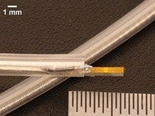
“So we’ve developed a dual-frequency intravascular ultrasound transducer which transmits and receives acoustic signals,” Jiang says. “Operating on two frequencies allows us to do everything the existing intravascular ultrasound devices can do, but also makes it much easier for us to detect the contrast agents – or microbubbles – used for molecular imaging and vasa vasorum detection.”
The prototype device has performed well in laboratory testing, but the researchers say they are continuing to optimize the technology. They hope to launch pre-clinical studies in the near future.
The Latest on: Detecting Risk for Heart Attack Stroke
[google_news title=”” keyword=”Detecting Risk for Heart Attack Stroke” num_posts=”10″ blurb_length=”0″ show_thumb=”left”]
via Google News
The Latest on: Detecting Risk for Heart Attack Stroke
- Alzheimer’s, Cancer To Diabetes: Important Tests To Get Depending On Your Family Historyon April 27, 2024 at 3:49 am
A family history of chronic diseases can put healthy individuals at risk Therefore getting regular tests is crucial for early diagnosis ...
- Use of antipsychotic meds for older adults drops after warning letter from Medicareon April 26, 2024 at 9:13 am
Warning letters sent by Medicare officials can prompt a decline in antipsychotic prescriptions for seniors with dementia, a new study finds.
- Harvard study reveals how pollution raises risk of heart attack or strokeon April 26, 2024 at 8:09 am
Air pollution raises the risk of a fatal heart attack or stroke in middle age by triggering stress, warns new research. The worrying findings suggest that "dirty air" puts under-65s in greater danger ...
- Rare condition that stunts growth also reduces heart attack riskon April 25, 2024 at 5:00 pm
People with an ultra-rare condition that stunts growth are less likely to suffer a heart attack or stroke, suggests new research.
- 5 women’s health tips to prevent and detect strokes, according to cardiologistson April 25, 2024 at 2:30 am
One in five women between the ages of 55 and 75 will experience a stroke. Parag Shah, MD, a physical medicine specialist in Jacksonville, Florida, shares tips for women to reduce their risk.
- AFib May be More Common in People Under 65 Than Previously Thoughton April 23, 2024 at 6:28 am
A new study suggests that atrial fibrillation (AFib) may be more common in people under 65 than shown by previous research ...
- Spouse’s stroke could boost partner’s depression risk, study suggestson April 20, 2024 at 3:30 am
Those whose spouses had a stroke or heart failure were at higher risk than those whose spouses had a heart attack.
- What cholesterol levels could lead to a heart attackon April 18, 2024 at 7:52 am
Cholesterol. It's a term often whispered with concern, leaving many wondering if their numbers spell impending doom. But fear not! Understanding chole ...
- Heart Failure, Not Stroke, Is Most Common Complication of AFib, Study Findson April 17, 2024 at 5:00 pm
A new study found that for those who receive a diagnosis of atrial fibrillation, the most common complication is heart failure, followed by stroke.
- Dangerous heart conditions often go undetected in pregnant and postpartum women even years later | Health Smarton April 16, 2024 at 3:40 am
National Jewish Health experts advocate for screenings to detect heart conditions that may develop in otherwise healthy women.
via Bing News
