


Fast fMRI tracks brain activity during human thought for first time
By significantly increasing the speed of functional MRI (fMRI), NIBIB-funded researchers have been able to image rapidly fluctuating brain activity during human thought. fMRI measures changes in blood oxygenation, which were previously thought to be too slow to detect the subtle neuronal activity associated with higher order brain functions. The new discovery that fast fMRI can detect rapid brain oscillations is a significant step towards realizing a central goal of neuroscience research: mapping the brain networks responsible for human cognitive functions such as perception, attention, and awareness.
“A critical aim of the President’s BRAIN Initiative1 is to move neuroscience into a new realm where we can identify and track functioning neural networks non-invasively,” explains Guoying Liu, Ph.D., Director of the NIBIB program in Magnetic Resonance Imaging. “This work demonstrates the potential of fMRI for mapping healthy neural networks as well as those that may contribute to neurological diseases such as dementia and other mental health disorders, which are significant national and global health problems.”

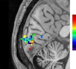
Oscillating checkerboards with different levels of contrast were sequentially presented to individuals to stimulate the visual cortex. Source: Lewis, et al.2
fMRI works by detecting local increases in oxygen as blood is delivered to a working part of the brain. The technique has been instrumental for identifying which areas in the brain control functions such as vision, hearing, or touch. However, standard fMRI can only detect the blood flow coming to replenish an area of the brain several seconds after it has performed a function. It was generally accepted that this was the limit of what could be detected by fMRI—identification of a region in the brain that had responded to a large stimulus, such as a continuous 30 second “blast” of bright light.
Combining several new techniques, Jonathan R. Polimeni, Ph.D., senior author of the study, and his colleagues at Harvard’s Athinoula A. Martinos Center for Biomedical Imaging, applied fast fMRI in an effort to track neuronal networks that control human thought processes, and found that they could now measure rapidly oscillating brain activity. The results of this groundbreaking work are reported in the October 2016 issue of the Proceedings of the National Academy of Sciences.2
The researchers used fast fMRI in human volunteers observing a rapidly fluctuating checkerboard pattern. The fast fMRI was able to detect the subtle and very rapid oscillations in cerebral blood flow in the brain’s visual cortex as the volunteers observed the changing pattern.
“The oscillating checkerboard pattern is a more “naturalistic” stimulus, in that its timing is similar to the very subtle neural oscillations made during normal thought processes,” explains Polimeni. “The fast fMRI detects the induced neural oscillations that allow the brain to understand what the eye is observing — the changing checkerboard pattern. These subtle oscillations were completely undetectable with standard fMRI. This exciting result opens the possibility of using fast fMRI to image neural networks as they guide the process of human thought.”
One such possibility is suggested by first author of the study Laura D. Lewis, Ph.D. “This technique now gives us a method for obtaining much more detailed information about the complex brain activity that takes place during sleep, as well as other dynamic switches in brain states, such as when under anesthesia and during hallucinations.”
Concludes Polimeni, “It had always been thought that fMRI had the potential to play a major role in these types of studies. Meaningful progress in cognitive neuroscience depends on mapping patterns of brain activity, which are constantly and rapidly changing with every experience we have. Thus, we are extremely excited to see our work contribute significantly to achieving this goal.”
Learn more: Imaging technique can see you think
The Latest on: Brain activity during human thought
[google_news title=”” keyword=”brain activity during human thought” num_posts=”10″ blurb_length=”0″ show_thumb=”left”]
via Google News
The Latest on: Brain activity during human thought
- Super-detailed map of brain cells that keep us awake could improve our understanding of consciousnesson May 1, 2024 at 11:50 am
It's a tool that could help scientists pinpoint which brain activity is "necessary and sufficient" to ... Related: What happens in our brains when we 'hear' our own thoughts? Human consciousness ...
- Elon Musk’s Neuralink begins clinical trials in Phoenixon May 1, 2024 at 4:59 am
Neuralink chose Barrow as the site for its PRIME Study based on the institute’s expertise for treating patients with complex neurological conditions. Barrow has more than 300 clinical trials underway ...
- Scientists restore brain cells impaired by a rare genetic disorderon April 30, 2024 at 3:01 am
A therapy that restores brain cells impaired by a rare genetic disorder may offer a strategy for treating conditions like autism, epilepsy, and schizophrenia.
- Flexible Microdisplay Monitors Brain Activity in Real-Time during Brain Surgeryon April 24, 2024 at 5:00 pm
A thin film that combines an electrode grid and LEDs can both track and produce a visual representation of the brain’s activity in real time during surgery—a huge ... Dayeh’s team pioneered human ...
- A flexible microdisplay that can monitor brain activity in real-time during brain surgeryon April 23, 2024 at 5:00 pm
Researchers have created a thin film that combines an electrode grid and LEDs that can both track and produce a visual representation of the brain's activity in real-time during surgery—a huge ...
- A flexible microdisplay can monitor brain activity in real-time during brain surgeryon April 23, 2024 at 5:00 pm
A thin film that combines an electrode grid and LEDs can both track and produce a visual representation of the brain's activity in real-time during surgery ... pioneered human brain and spinal ...
- Exploring brain synchronization patterns during social interactionson April 22, 2024 at 4:59 pm
Social interactions synchronize brain activity ... networking during social interactions," says lead researcher Dr. Yuto Kurihara, Research Associate at the Faculty of Human Sciences at Waseda ...
- When thoughts flow in one directionon April 18, 2024 at 11:38 am
Contrary to previous assumptions, nerve cells in the human neocortex are wired differently than in mice. Those are the findings of a new study conducted by Charité – Universitätsmedizin Berlin and ...
- When technology can read your brain waves, who owns your thoughts?on April 18, 2024 at 10:34 am
By Mack DeGeurin | Published Apr 18, 2024 1:35 PM EDT Brain Computer Interface (BCI) companies are charging ahead with devices and services attempting to understand and manipulate human neural ...
via Bing News

