


A camera makes hidden tumors visible during an operation
Cancer patients have the highest probability of recovering if tumors are completely removed. However, tiny clusters of cancer cells are often difficult for surgeons to recognize and remove. A camera makes hidden tumors visible during an operation.
Tumor removal surgeries pose a great challenge even to skillful and experienced surgeons. For one thing, tumor margins are blending into healthy tissue and are difficult to differentiate. For another, distributed domains of cancer and pre-malignancies are difficult to recognize. Up to now, doctors depend exclusively upon their trained eyes when excising pieces of tumors. In future, a new special camera system can help visualize during operation even the smallest, easy-to-overlook malignant pieces of tumor and thereby support the surgeons during complicated interventions.
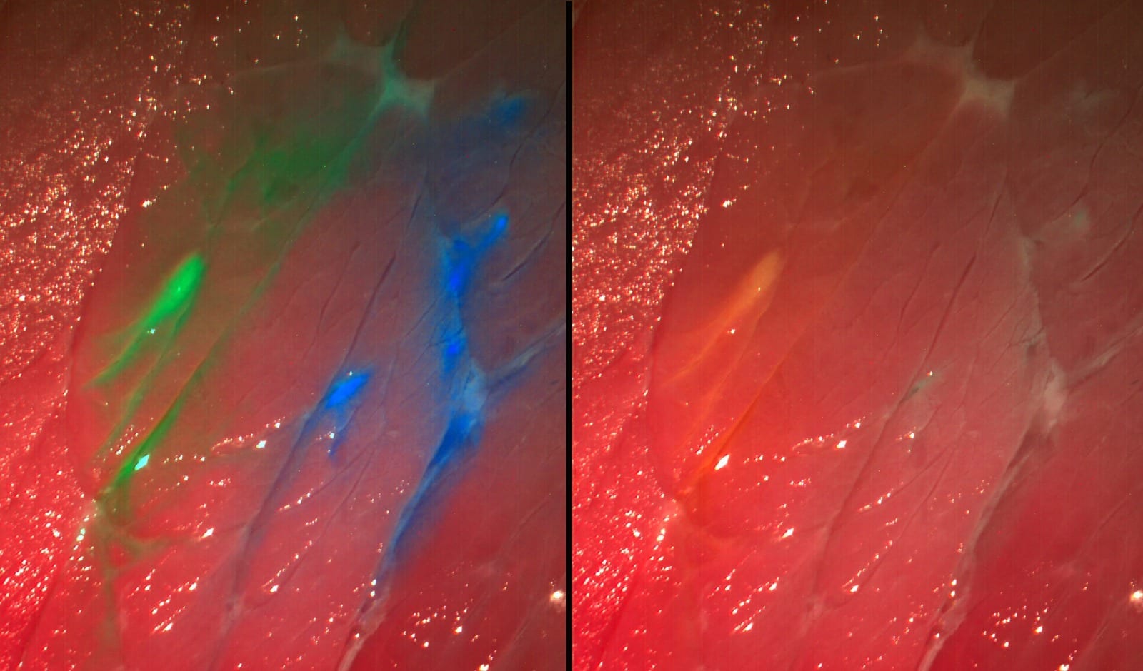
The trick: the camera can display fluorescent molecules that “paint” the cancer tissue. These are injected into the patients blood circulation prior to the operation and selectively attach onto the tumor during their trip through the body. If the corresponding area is then illuminated with a specific wavelength, fluorescence is emitted and the malignant tissue glows green, blue, red, or any other color, depending on the injected dye, while the healthy tissue appears the same. In this way, the surgeon can see clusters of tumors cells that cannot be recognized by the naked eye.
New system reveals several dyes simultaneously
Researchers at the Fraunhofer Project Group for Automation in Medicine and Biotechnology (PAMB), which belongs to the Fraunhofer Institute for Manufacturing Engineering and Automation (IPA), have developed a new surgical aid, a multispectral fluorescence camera system. In the future, this special camera will integrate into various medical imaging systems such as, surgical microscopes and endoscopes, etc. The scientists from Mannheim, Germany, will make the debut of a prototype of this high-tech system at the Medica Trade Fair in Düsseldorf in the joint Fraunhofer booth (Halle 10, Booth F05) between 20-23 November. The novel aspect about this camera: it can display several fluorescent dyes and the reflectance image simultaneously in real time – systems available until now have not been able to achieve this. The advantage: arteries and delicate nerves that must not be injured during an intervention can likewise be colored with dye. They too can then be detected with the new camera, since they are set apart from their surroundings.
“The visibility of the dye to the camera depends in large part on the selection of the correct set of fluorescence filters. The filter separates the incident excitation wave- lengths from the fluorescing wavelengths so that the diseased tissue is also set apart from its surroundings, even at very low light intensities,” says Nikolas Dimitriadis, a scientist at PAMB. The researchers and their colleague require only one camera and one set of filters for their photographs, which can present up to four dyes at the same time. Software developed in-house analyses and processes the images in seconds and presents it continuously on a monitor during surgery. The information from the fluorescent image is superposed on the normal color image. “The operator receives significantly more accurate information. Millimeter-sized tumor remnants or metastases that a surgeon would otherwise possibly overlook are recognizable in detail on the monitor. Patients operated under fluorescent light have improved chances of survival,” says Dr. Nikolaos Deliolanis, head of the Biomedical Optics Group at PAMB.
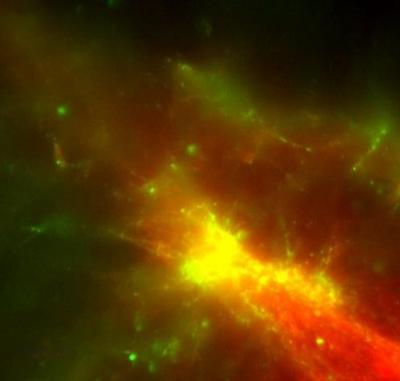
In order to be able to employ the multispectral fluorescence camera system as adapt- ably as possible, it can be converted to other combinations of dyes. “One preparation that is already available to make tumors visible is 5-amino levulinic acid (5-ALA). Physicians employ this especially for glioblastomas – one of the most frequent malignant brain tumors in adults,” explains Dimitriadis. 5-ALA leads to an accumulation of a red dye in the tumor and can likewise be detected with the camera. The multispectral fluorescence imaging system should have passed testing for use with humans as soon as next year.
Go deeper with Bing News on:
Cancer detection
- Prince William gives rare health update about Princess Kate amid her cancer diagnosis
Prince William is providing a royal update about wife Princess Kate's cancer diagnosis two months after she announced her health news to the world.
- Queen Camilla Wears Fiona Clare Leopard-print Dress With King Charles III for Charity Cancer Research Hospital Visit
For the special occasion, Queen Camilla fashioned a Fiona Clare dress, which featured a leopard print design in shades of muted green, black, white and brown. The dress also featured ruffled details on the bodice.
Go deeper with Google Headlines on:
Cancer detection
[google_news title=”” keyword=”cancer detection” num_posts=”5″ blurb_length=”0″ show_thumb=”left”]
Go deeper with Bing News on:
Multispectral fluorescence camera
- Feed has no items.
Go deeper with Google Headlines on:
Multispectral fluorescence camera
[google_news title=”” keyword=”multispectral fluorescence camera” num_posts=”5″ blurb_length=”0″ show_thumb=”left”]
