


A research team led by UC Berkeley engineers has developed a new smartphone microscope that uses video to automatically detect and quantify infection by parasitic worms in a drop of blood. This next generation of UC Berkeley’s CellScope technology could help revive efforts to eradicate debilitating filarial diseases in Africa by providing critical information to health providers in the field.
“We previously showed that mobile phones can be used for microscopy, but this is the first device that combines the imaging technology with hardware and software automation to create a complete diagnostic solution,” said Daniel Fletcher, an associate chair and professor of bioengineering, whose UC Berkeley lab pioneered the CellScope. “The video CellScope provides accurate, fast results that enable health workers to make potentially life-saving treatment decisions in the field.”
The UC Berkeley engineers teamed up with Dr. Thomas Nutman from the National Institute of Allergy and Infectious Diseases (NIAID), and collaborators from Cameroon and France to develop the device. They conducted a pilot study in Cameroon, where health officials have been battling the parasitic worm diseases onchocerciasis (river blindness) and lymphatic filariasis.
The video CellScope, which uses motion instead of molecular markers or fluorescent stains to detect the movement of worms, was as accurate as conventional screening methods, the researchers found. The results of the pilot study are reported today (Wednesday, May 6) in the journal Science Translational Medicine.
“This research is addressing neglected tropical diseases,” said Fletcher. “It demonstrates what technology can do to help fill a void for populations that are suffering from terrible, but treatable, diseases.”
Battling parasitic worms
River blindness is transmitted through the bite of blackflies and is the second-leading cause of infectious blindness worldwide. Lymphatic filariasis, spread by mosquitoes, leads to elephantiasis, a condition marked by painful, disfiguring swelling. It is the second-leading cause of disability worldwide and, like river blindness, is highly endemic in certain regions in Africa.
The antiparasitic drug ivermectin, or IVM, can be used to treat these diseases, but mass public health campaigns to administer the medication have been stalled because of potentially fatal side effects for patients co-infected with Loa loa, which causes loiasis, or African eye worm. When there are high circulating levels of microscopic Loa loaworms in a patient, treatment with IVM can potentially lead to severe or fatal brain or other neurologic damage.
The standard method of screening for levels of Loa loa involves trained technicians manually counting the worms in a blood smear using conventional laboratory microscopes, making the process impractical for use in field settings and in mass campaigns to administer IVM.
The serious side effects of Loa loa and the difficulty of rapidly quantifying Loa levels in patients before treatment make it too risky to broadly administer IVM, representing a major setback in the efforts to eradicate river blindness and elephantiasis.
Next generation CellScope uses video, automation
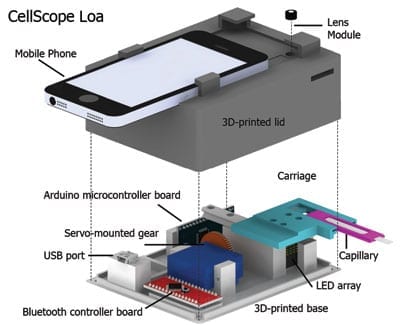
For this latest generation of the mobile phone microscope, named CellScope Loa, the researchers paired a smartphone with a 3D-printed plastic base where the sample of blood is positioned. The base included LED lights, microcontrollers, gears, circuitry and a USB port.
Control of the device is automated through an app the researchers developed for this purpose. With a single touch of the screen by the healthcare worker, the phone communicates wirelessly via Bluetooth to controllers in the base to process and analyze the sample of blood. Gears move the sample in front of the camera, and an algorithm automatically analyzes the telltale “wriggling” motion of the worms in video captured by the phone. The worm count is then displayed on the screen.
Fletcher said previous field tests revealed that automation helped reduce the rate of human error. The procedure takes about two minutes or less, starting from the time the sample is inserted to the display of the results. Pricking a finger and loading the blood onto the capillary adds another minute to the time.
The short processing time allows health workers to quickly determine on site whether it is safe to administer IVM.

Read more: Smartphone video microscope automates detection of parasites in blood
The Latest on: Smartphone microscope
[google_news title=”” keyword=”Smartphone microscope” num_posts=”10″ blurb_length=”0″ show_thumb=”left”]
via Google News
The Latest on: Smartphone microscope
- With contactless screening tech, this Toronto startup hopes to catch breast cancer early — and save liveson May 9, 2024 at 5:00 am
Amid evidence of rising breast cancer rates among young women in Canada, one Toronto startup is offering a contactless and radiation-free device that can help doctors identify suspicious changes in ...
- The 7 best cell phones of 2024 go way, way beyond talking and textingon April 18, 2024 at 11:01 am
The latest smartphone can handle so much more than just calls or texts. They're powerful communications, productivity, fitness and entertainment devices -- plus so much more. But that also means ...
- The Basics of Smartphone Backupson April 16, 2024 at 5:01 pm
A backup saves file copies at a certain point in time. Syncing your smartphone keeps information in certain apps, like contacts and calendars, current across multiple devices. When synchronized ...
- Social psychologist urges parents to keep smartphones away from kids to 'protect' their mental healthon April 11, 2024 at 1:00 pm
Smartphones are creating an anxiety and depression-riddled generation of teens, according to social psychologist and New York University professor Jonathan Haidt, but there are steps parents can ...
- 10 Best Gosky Microscopeson April 10, 2024 at 5:00 pm
[High Magnification] WF10X WF25X eyepiece alone or with 2X lens, cooperate 4x 10x 40x Achromatic Objectives lenses, biological microscopes offers10 magnification settings, 40x, 80x, 100x ...
- 10 Best Handheld Microscope For Cannabison April 10, 2024 at 5:00 pm
Huge Variety - The illuminated loupe is great for just about anything that needs some zoom, coin magnifying glass with light, grow microscope, diamond magnifying glass, Jewelry loop magnifier and more ...
- The best microscopes for students in 2024on April 10, 2024 at 1:00 pm
Here are the microscopes to help them get started. This classic microscope is made with students’ needs in mind. It can connect to a laptop or other screen for group viewing, helping large ...
- Under the Microscope: Can Apple Stock Thrive Under Regulatory Scrutiny?on April 3, 2024 at 3:35 am
The Justice Dept. and 16 states are going after Apple stock for allegedly maintaining an illegal monopoly in the smartphone market. You know the thing that makes users value Apple’s ecosystem so ...
- Exploring the Unseen The Best Microscopes for Kids in 2024on March 4, 2024 at 2:32 am
Popular Science Smartphone Microscope (Age 6+): Embrace the power of technology with the Popular Science Smartphone Microscope, suitable for children aged 6 and above. By harnessing the camera ...
- Characteristics of a Smartphoneon February 9, 2024 at 6:00 am
Today's smartphones are able to text message, access the Internet, exchange email, take videos and pictures, update Facebook and Twitter statuses and more. While the aforementioned are a few ...
via Bing News

