


Image: Courtesy of the Salk Institute for Biological Studies
New technique to selectively and noninvasively turn on groups of neurons in worms could be boon to science and medicine
Salk scientists have developed a new way to selectively activate brain, heart, muscle and other cells using ultrasonic waves. The new technique, dubbed sonogenetics, has some similarities to the burgeoning use of light to activate cells in order to better understand the brain.
This new method–which uses the same type of waves used in medical sonograms–may have advantages over the light-based approach–known as optogenetics–particularly when it comes to adapting the technology to human therapeutics. It was described September 15, 2015 in the journal Nature Communications.
“Light-based techniques are great for some uses and I think we’re going to continue to see developments on that front,” says Sreekanth Chalasani, an assistant professor in Salk’s Molecular Neurobiology Laboratory and senior author of the study. “But this is a new, additional tool to manipulate neurons and other cells in the body.”
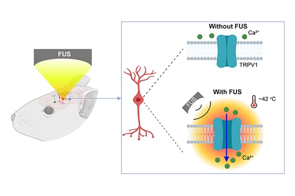
In optogenetics, researchers add light-sensitive channel proteins to neurons they wish to study. By shining a focused laser on the cells, they can selectively open these channels, either activating or silencing the target neurons. But using an optogenetics approach on cells deep in the brain is difficult: typically, researchers have to perform surgery to implant a fiber optic cable that can reach the cells. Plus, light is scattered by the brain and by other tissues in the body.
Chalasani and his group decided to see if they could develop an approach that instead relied on ultrasound waves for the activation. “In contrast to light, low-frequency ultrasound can travel through the body without any scattering,” he says. “This could be a big advantage when you want to stimulate a region deep in the brain without affecting other regions,” adds Stuart Ibsen, a postdoctoral fellow in the Chalasani lab and first author of the new work.
Chalasani and his colleagues first showed that, in the nematodeCaenorhabditis elegans, microbubbles of gas outside of the worm were necessary to amplify the low-intensity ultrasound waves. “The microbubbles grow and shrink in tune with the ultrasound pressure waves,” Ibsen says. “These oscillations can then propagate noninvasively into the worm.”
Next, they found a membrane ion channel, TRP-4, which can respond to these waves. When mechanical deformations from the ultrasound hitting gas bubbles propagate into the worm, they cause TRP-4 channels to open up and activate the cell. Armed with that knowledge, the team tried adding the TRP-4 channel to neurons that don’t normally have it. With this approach, they successfully activated neurons that don’t usually react to ultrasound.
Read more: Salk scientists use sound waves to control brain cells
The Latest on: Sonogenetics
[google_news title=”” keyword=”sonogenetics” num_posts=”10″ blurb_length=”0″ show_thumb=”left”]
via Google News
The Latest on: Sonogenetics
- RWTH: Professor Andreas Herrmann Awarded ERC Advanced Granton April 19, 2024 at 9:56 pm
The Scientific Director of the DWI – Leibniz Institute for Interactive Materials is developing a new technology platform for controlling the activity of genes, proteins, and active pharmaceutical ...
- ERC Advanced Grant: Andreas Herrmann receives 2.5 million euros from the EUon April 15, 2024 at 11:42 pm
as part of the ERC Advanced Grant for his project "Remote controlling biological systems by sonopharmacology and sonogenetics" (SONOPHARMAGEN). Andreas Herrmann has thus succeeded in acquiring his ...
via Bing News