


The field of medicine is always on the lookout for better disease diagnostic tools—simpler, faster, and cheaper technologies to enhance patient treatment and outcomes.
Currently, microfluidic bioassay devices are the preferred diagnostic tools that allow clinicians to measure the concentration of disease biomarkers within a patient’s biological sample, such as blood. They can indicate the likelihood of a disease based on a comparison of the biomarker concentration in the sample relative to the normal level. To detect this concentration, the patient’s sample is passed across a surface containing immobilized bioreceptors, or “biomarker-capturing” molecules that have been attached to this surface. A researcher can then record the biomarker abundance, determine whether the level is normal, and reach a diagnosis. Since the efficiency of these devices relies on how intact and functional the attached bioreceptors are, immobilizing these bioreceptors without causing damage has proved daunting.
Over the last two decades, microcontact printing, which uses a rubber stamp to immobilize the bioreceptors, has been established as a robust method to create a variety of assays with multiple applications. Yet this method also has its flaws, particularly when utilized at the nano scale—the scale where proteins and DNA reign. At this scale, the harsh and elaborate techniques currently used compromise the device’s resolution, whether by deforming the stamp or damaging the bioreceptors, thus yielding data somewhat unmanageable for use in diagnostics or other applications. However, in a recent article published in the journal Analyst, researchers at the Okinawa Institute of Science and Technology Graduate University (OIST) describe a new sequence of printing steps that have rectified these issues.
For microcontact printing, “you need a stamp, an ink, and a surface, and then you create your pattern on your surface. It’s as simple as that,” explains Shivani Sathish, OIST PhD student in the Micro/Bio/Nanofluidics Unit, and first author on the paper.
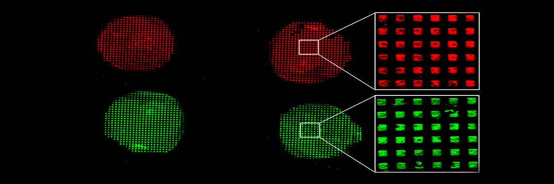
The stamp is made of polydimethylsiloxane, which is a flexible solid similar to the rubber used in everyday stamps. The ink is a solution composed of silicon- and oxide-containing molecules called APTES, and the surface is glass. After coating the stamp with the ink, the stamp is pressed onto the glass, and then removed after a short incubation. The result is a patterned layer of APTES on the glass—a checkerboard of regions with or without APTES (Figure, ii). Next, a microfluidic device, which contains one or more microchannels configured to guide fluid through specified pathways, is sealed over the patterned glass (Figure, iv). Finally, the bioreceptors are chemically linked to the APTES regions within the microfluidic channels. The device as a whole is about the size of a postage stamp.
The system is now ready for use as a diagnostic assay. To carry out the assay, a fluid sample from a patient is delivered through the microfluidic device attached to the glass. If the pertinent disease biomarker is present, the molecule will “stick” to the areas containing the bioreceptors.
What is important about the APTES solution is its convenient chemistry. “Depending on your bioreceptor of interest, you just have to choose the appropriate chemistry to link the molecule with the APTES,” Ms. Sathish explains. Or in other words, one stamp can be used to prepare an assay with the ability to immobilize a variety of different bioreceptors—one stamp allows for multiple tests and diagnoses on a single surface. This feature would be advantageous for diagnosing complex diseases such as cancer, which relies on tests that can detect multiple markers to improve the diagnosis.
In their research, Ms. Sathish and colleagues developed an improved technique to create the most optimal disease diagnostic device for use at the nano scale. Here, they first patterned nanoscale features of APTES using an ink made of APTES in water, as opposed to harsh chemicals, which eliminated the stamp-swelling issue. Then, they immobilized the bioreceptors onto the surface as the very last step of the process, after patterning the APTES and attaching the microfluidic device. By attaching the bioreceptors as the final step, the researchers avoided exposing them to extreme and damaging conditions. They then demonstrated the efficacy of the final device by running an assay to capture the biomarkers interleukin 6 and human c-reactive protein, two substances that are often elevated in the body during inflammation.
“The final goal is to create a point-of-care device,” explains OIST Professor Amy Shen, who headed the research.
“If you get your bioreceptors pre-immobilized within microfluidic devices you can then use them as diagnostic tools as and when required,” Ms. Sathish continues. “[Eventually] instead of having a whole clinical team that processes your sample…we’re hoping that the patients can do it themselves at home.”
Learn more: Miniature Technology, Big Hope for Disease Detection
The Latest on: Disease detection
[google_news title=”” keyword=”disease detection” num_posts=”10″ blurb_length=”0″ show_thumb=”left”]- How to stop kidney disease from getting worse?on April 27, 2024 at 2:30 pm
Kidney diseases pose a global health threat. Preventive measures include a healthy lifestyle, routine check-ups, and avoiding unnecessary medications.
- Catanduanes farmers get training in identifying abaca diseases, viruses to help sustain the industryon April 27, 2024 at 9:04 am
Abaca farmers from Catanduanes found renewed hope through a training program bout the plant’s disease and virus detection. Twenty-one farmers attended the training organized by the Department of ...
- Chronic wasting disease detected in Edwards County deer breeding facilityon April 25, 2024 at 11:24 am
The Odessa American is the leading source of local news, information, entertainment and sports for the Permian Basin.
- Fatal ‘zombie deer’ disease found in Harpers Ferry National Historical Parkon April 24, 2024 at 12:20 pm
Two white-tailed deer inside Harpers Ferry National Historical Park in West Virginia have tested positive for the fatal Chronic Wasting Disease.
- Fatal 'zombie deer' disease found in West Virginia national historic parkon April 24, 2024 at 8:06 am
Two white-tailed deer inside Harpers Ferry National Historical Park in West Virginia have tested positive for the fatal Chronic Wasting Disease, the National Park Service announced Tuesday.
- Company developing early cancer-detection tests to cut 20% of workforceon April 24, 2024 at 7:35 am
Early cancer-detection test maker that raised $254 million earlier this year now says it will cut more than 100 jobs — about 20% of its workforce — as it restructures ...
- 2024 Report | European Biopsy Devices Market to Reach US$ 1,074.0 Million by 2032: Enhanced Focus on Early Disease Detection Fuels Growthon April 24, 2024 at 6:41 am
It is projected to grow at a CAGR of 5.99% from 2024 to 2032. The analyst forecasts it to reach US$ 1,074.0 Million by 2032. The increasing emphasi ...
- Researchers aim to use AI for early screening and prognosis of Dry Eye Diseaseon April 23, 2024 at 1:19 pm
Dry Eye Disease (DED) is one of the more common eye diseases, affecting up to 30% of the world's population. This disease can affect many different types of people and can wind up being a great ...
- Artificial intelligence to be used for the detection of common eye diseaseon April 23, 2024 at 6:14 am
Here is where the combined efforts of ophthalmic disease detection and the world of computer scientists and engineers can help. “By addressing challenges, imparting insights, and delineating future ...
- Women's heart disease is underdiagnosed, but new machine learning models can help solve this problemon April 22, 2024 at 9:00 pm
When it comes to matters of the heart, cardiovascular disease in women is underdiagnosed compared to men. A popular scoring system used to estimate how likely a person is to develop a cardiovascular ...
via Google News and Bing News
