CREDIT
Adapted from Viola et al., Nature Nanotechnology, 2014.
MRI probe technology shows brain toxins in living animals for first time
No methods currently exist for the early detection of Alzheimer’s disease, which affects one out of nine people over the age of 65. Now, an interdisciplinary team of Northwestern University scientists and engineers has developed a noninvasive MRI approach that can detect the disease in a living animal. And it can do so at the earliest stages of the disease, well before typical Alzheimer’s symptoms appear.
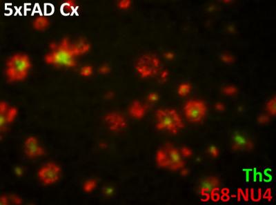
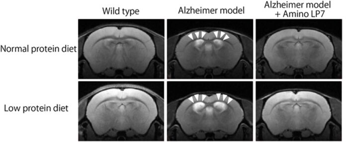
Led by neuroscientist William L. Klein and materials scientist Vinayak P. Dravid, the research team developed an MRI (magnetic resonance imaging) probe that pairs a magnetic nanostructure (MNS) with an antibody that seeks out the amyloid beta brain toxins responsible for onset of the disease. The accumulated toxins, because of the associated magnetic nanostructures, show up as dark areas in MRI scans of the brain.
This ability to detect the molecular toxins may one day enable scientists to both spot trouble early and better design drugs or therapies to combat and monitor the disease. And, while not the focus of the study, early evidence suggests the MRI probe improves memory, too, by binding to the toxins to render them “handcuffed” to do further damage.
“We have a new brain imaging method that can detect the toxin that leads to Alzheimer’s disease,” said Klein, who first identified the amyloid beta oligomer in 1998. He is a professor of neurobiology in the Weinberg College of Arts and Sciences.
Take me to the story: New non-invasive method can detect Alzheimer’s disease early