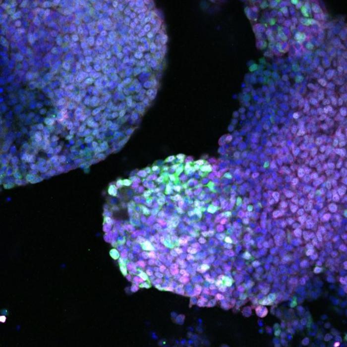
In some cases, looking at a living cell under a microscope can cause it damage or worse, can kill it. Now, a new kind of microscope has been invented by researchers from the Howard Hughes Medical Institute that is able to non-invasively take a three dimensional look inside living cells with stunning results. The device uses a thin sheet of light like that used to scan supermarket bar codes and could help biologists to achieve their goal of understanding the rules that govern molecular processes within a cell.
Veteran microscope innovator Eric Betzig says that the field of microscopy has been hindered by the fact that many techniques require cells to be killed and fixed before being viewed. Light produced by microscopes used for live-cell techniques can, in some cases, actually cause damage to the cells. The light also floods the whole area being examined, not just the small portion that’s in focus – producing blur from the out-of-focus regions.
Two years after arriving at HHMI’s Janelia Farm Research Campus, Betzig started working ways to overcome these problems.
“The question was, is there a way of minimizing the amount of damage you’re doing so that you can then study cells in a physiological manner while also studying them at high spatial and temporal resolution for a long time?” said Betzig.


