


When women undergo lumpectomies to remove breast cancer, doctors try to remove all the cancerous tissue while conserving as much of the healthy breast tissue as possible.
But currently there’s no reliable way to determine during surgery whether the excised tissue is completely cancer-free at its margins — the proof that doctors need to be confident that they removed all of the tumor. It can take several days for pathologists using conventional methods to process and analyze the tissue.
That’s why between 20 and 40 percent of women have to undergo second, third or even fourth breast-conserving surgeries to remove cancerous cells that were missed during the initial procedure, according to studies.
A new microscope invented by a team of University of Washington mechanical engineers and pathologists could help solve this, and other, problems. It can rapidly and non-destructively image the margins of large fresh tissue specimens with the same level of detail as traditional pathology — in no more than 30 minutes.
“Surgeons are sort of flying blind during these breast-conserving surgeries,” said mechanical engineering professor Jonathan Liu. “Oftentimes they’ve left some tumor behind which they don’t know about until a few days later when the pathologist finds it.”
“If we can rapidly image the entire surface or margin of the excised tissue during the procedure, we can tell them if they still have tumor left in the body or not. And that would be a huge benefit to cancer patients,” Liu said.

UW Medicine professor of pathology Larry True (left) and mechanical engineering associate professor Jonathan Liu (right) led the team that developed the light-sheet microscope and demonstrated its utility for various clinical applications.Mark Stone/University of Washington
The new light-sheet microscope — which is described in a new paper published June 26 in Nature Biomedical Engineering — offers other advantages over existing processes and microscope technologies. It conserves valuable tissue for genetic testing and diagnosis, quickly and accurately images the irregular surfaces of large clinical specimens, and allows pathologists to zoom in and “see” biopsy samples in three dimensions.
“The tools we use in pathology have changed little over the past century,” said co-author Nicholas Reder, chief resident and clinical research fellow in UW Medicine’s Department of Pathology. “This light-sheet microscope represents a major advance for pathology and cancer patients, allowing us to examine tissue in minutes rather than days and to view it in three dimensions instead of two — which will ultimately lead to improved clinical care.”
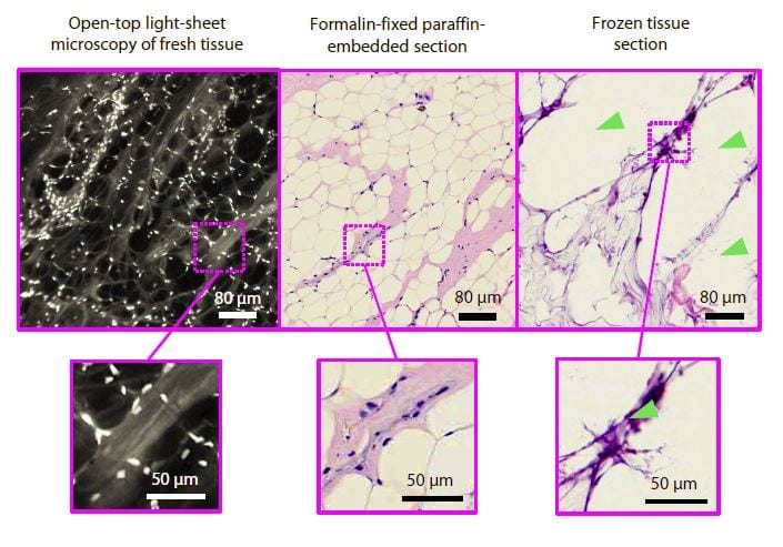
Current pathology techniques involve processing and staining tissue samples, embedding them in wax blocks, slicing them thinly, mounting them on slides, staining them, and then viewing these two-dimensional tissue sections with traditional microscopes — a process that can take days to yield results.
Another technique to provide real-time information during surgeries involves freezing and slicing the tissue for quick viewing. But the quality of those images is inconsistent, and certain fatty tissues, such as those from the breast, do not freeze well enough to reliably use the technique.
By contrast, the UW open-top light-sheet microscope uses a sheet of light to optically “slice” through and image a tissue sample without destroying any of it. All of the tissue is conserved for potential downstream molecular testing, which can yield additional valuable information about the nature of the cancer and lead to more effective treatment decisions.
“Slide-based pathology is still an analog technique, much like radiology was several decades ago when X-rays were obtained on film. By imaging tissues in 3-D without having to mount thin tissue sections on glass slides, we are trying to transform pathology much like 3-D X-ray CT has transformed radiology,” Liu said. “While it is possible to scan microscope slides for digital pathology, we digitally image the intact tissues and bypass the need to prepare slides, which is simpler, faster and potentially less expensive.”

UW Medicine pathologists Larry True (left) and Nicholas Reder (right) prepare a tissue sample for imaging on the light-sheet microscope. No tissue is destroyed in the staining and imaging process, which preserves that valuable resource for downstream molecular testing.Mark Stone/University of Washington
“If we can do this without consuming any tissue, so much the better,” said co-author Larry True, professor of pathology at UW Medicine. “We want to use that valuable tissue for purposes which are becoming ever more important for treating patients — such as sequencing the tumor cells and finding genetic abnormalities that we can target with specific drugs and other precision medicine techniques.”
The light-sheet microscope also offers advantages over other non-destructive optical- sectioning microscopes on the market today, which process images slowly and have difficulty maintaining the optimal focus when dealing with clinical specimens, which always have microscopic surface irregularities.
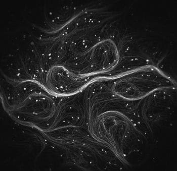
The UW microscope can both image large tissue surfaces at high resolution and stitch together thousands of two-dimensional images per second to quickly create a 3-D image of a surgical or biopsy specimen. That additional data could one day allow pathologists to more accurately and consistently diagnose and grade tumors.
“Pathologists are currently very limited in how much they can look at on a glass slide,” said co-author Adam Glaser, a postdoctoral fellow in the UW Molecular Biophotonics Laboratory. “If we can give them three-dimensional data, we can give them more information to help improve the accuracy of a patient’s diagnosis.”

Mechanical engineering postdoctoral fellow Adam Glaser assembles the next generation of the light-sheet microscope, which will provide greater resolving power and imaging depth than the first system.Mark Stone/University of Washington
The UW team achieved these improvements by configuring various optical technologies in new ways and optimizing them for clinical use. Their open-top arrangement, which places all of the optics underneath a glass plate, allows them to image larger tissues than other microscopes.
The team is currently working on speeding up the optical-clearing process that allows light to penetrate biopsy samples more easily. Future areas of research include optimizing their 3-D immunostaining processes, as well as as continuing a collaboration formed during the UW eScience Institute’s Winter Incubator program with Dr. Ariel Rokem to develop algorithms that can process the vast amounts of 3-D pathology data that their system generates, with the ultimate goal of helping pathologists zero in on suspicious areas of tissue.
Learn more: Microscope can scan tumors during surgery and examine cancer biopsies in 3-D
The Latest on: Light-sheet microscope
[google_news title=”” keyword=”light-sheet microscope” num_posts=”10″ blurb_length=”0″ show_thumb=”left”]- 30 Times Clearer – Scientists Develop Improved Mid-Infrared Microscopeon April 24, 2024 at 3:55 am
The chemical images taken of the insides of bacteria were 30 times clearer than those from conventional mid-infrared microscopes. Researchers at the University of Tokyo have developed an advanced ...
- Scientists Solve Decades-Old Microscopy Problemon April 24, 2024 at 1:55 am
Studying tissues, cells, and proteins under a microscope is essential for disease prevention and treatment. This research requires accurately measuring the dimensions of these biological structures.
- News tagged with prostate canceron April 14, 2024 at 5:00 pm
Researchers have incorporated a swept illumination source into an open-top light-sheet microscope to enable improved optical sectioning over a larger area of view. The advance makes the technique ...
- Researchers add swept illumination to open-top light-sheet microscopeon March 20, 2024 at 12:59 pm
The researchers used their improved open-top light-sheet microscope to capture images of densely labeled clinical specimens, showing its potential for nondestructive 3D pathology. Kevin W. Bishop ...
- Researchers add swept illumination to open-top light-sheet microscopeon March 20, 2024 at 12:06 pm
Researchers have incorporated a swept illumination source into an open-top light-sheet microscope to enable improved optical sectioning over a larger area of view. The advance makes the technique ...
- Investigating cells with a light microscopeon February 14, 2024 at 9:12 am
To use a light microscope to examine animal or plant cells. To make observations and draw scale diagrams of cells. Turn the coarse focus so that the stage is as close to the objective lens as ...
- UAB Institutional Research Core Programon December 16, 2023 at 1:17 pm
They are pictured with the Zeiss Lightsheet 7 light sheet microscope, an advanced imaging device capable of fluorescent imaging of living cells and tissues. What it does: A light sheet fluorescent ...
- Light Microscopy – Webinars and Online Eventson August 16, 2023 at 3:24 am
In this webinar, you'll learn about the principles behind light sheet microscopy, how it enhances analysis of complex biological systems and its diverse applications in neuroscience and cancer ...
via Google News and Bing News
