


Microsponges derived from seaweed may help diagnose heart disease, cancers, HIV and other diseases quickly and at far lower cost than current clinical methods.
The microsponges are an essential component of Rice University’s Programmable Bio-Nano-Chip (PBNC) and the focus of a new paper in the journal Small.
The paper by John McDevitt, the Brown-Wiess Professor in Bioengineering and Chemistry, and his colleagues at Rice’s BioScience Research Collaborative views the inner workings of PBNCs, which McDevitt envisions as a mainstream medical diagnostic tool.
PBNCs to diagnose a variety of diseases are currently the focus of six human clinical trials. McDevitt will discuss their development at the annual meeting of the American Association for the Advancement of Science (AAAS) in Washington, D.C., Feb. 17-21.
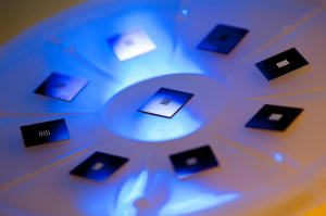
PBNCs capture biomarkers — molecules that offer information about a person’s health — found in blood, saliva and other bodily fluids. The biomarkers are sequestered in tiny sponges set into an array of inverted pyramid-shaped funnels in the microprocessor heart of the credit card-sized PBNC.
When a fluid sample is put into the disposable device, microfluidic channels direct it to the sponges, which are infused with antibodies that detect and capture specific biomarkers. Once captured, they can be analyzed within minutes with a sophisticated microscope and computer built into a portable, toaster-sized reader.
The biomarker capture process is the subject of the Smallpaper. The microsponges are 280-micrometer beads of agarose, a cheap, common, lab-friendly material derived from seaweed and often used as a matrix for growing live cells or capturing proteins.
The beauty of agarose is its ability to capture a wide range of targets from relatively huge protein biomarkers to tiny drug metabolites. In the lab, agarose starts as a powder, like Jell-O. When mixed with hot water, it can be formed into gels or solids of any size. The size of the pores and channels in agarose can be tuned down to the nanoscale.
The challenge, McDevitt said, was defining a new concept to quickly and efficiently capture and detect biomarkers within a microfluidic circuit. The solution developed at Rice is a network of microsponges with tailored pore sizes and nano-nets of agarose fibers. The sponge-like quality allows a lot of fluid to be processed quickly, while the nano-net provides a huge surface area that can be used to generate optical signals 1,000 times greater than conventional refrigerator-sized devices. The mini-sensor ensembles, he said, pack maximum punch.
The team found that agarose beads with a diameter of about 280 micrometers are ideal for real-world applications and can be mass-produced in a cost-effective way. These agarose beads retain their efficiency at capturing biomarkers, are easy to handle and don’t require specialized optics to see.
McDevitt and his colleagues tested beads with pores up to 620 nanometers and down to 45 nanometers wide. (A sheet of paper is about 100,000 nanometers thick.) Pores near 140 nanometers proved best at letting proteins infuse the beads’ internal nano-nets quickly, a characteristic that enables PBNCs to test for disease in less than 15 minutes.
The team reported on experiments using two biomarkers, carcinoembryonic antigens and Interleukin-1 beta proteins (and matching antibodies for both), purchased by the lab. After soaking the beads in the antibody solutions, the researchers tested their ability to recognize and capture their matching biomarkers. In the best cases, they showed near-total efficiency (99.5 percent) in the detection of bead-bound biomarkers.
