


Images of prisoners’ brains show important differences between those who are diagnosed as psychopaths and those who aren’t, according to a new study led by University of Wisconsin-Madison researchers.
The results could help explain the callous and impulsive antisocial behavior exhibited by some psychopaths.
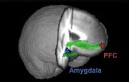
The study showed that psychopaths have reduced connections between the ventromedial prefrontal cortex (vmPFC), the part of the brain responsible for sentiments such as empathy and guilt, and the amygdala, which mediates fear and anxiety.
Two types of brain images were collected. Diffusion tensor images (DTI) showed reduced structural integrity in the white matter fibers connecting the two areas, while a second type of image that maps brain activity, a functional magnetic resonance image (fMRI), showed less coordinated activity between the vmPFC and the amygdala.
“This is the first study to show both structural and functional differences in the brains of people diagnosed with psychopathy,” says Michael Koenigs, assistant professor of psychiatry in the University of Wisconsin School of Medicine and Public Health. “Those two structures in the brain, which are believed to regulate emotion and social behavior, seem to not be communicating as they should.”
The study, which took place in a medium-security prison in Wisconsin, is a unique collaborative between three laboratories, UW-Madison psychology Professor Joseph Newman has had a long term interest in studying and diagnosing those with psychopathy and has worked extensively in the Wisconsin corrections system.
Dr. Kent Kiehl, of the University of New Mexico and the MIND Research Network, has a mobile MRI scanner that he brought to the prison and used to scan the prisoners’ brains. Koenigs and his graduate student, Julian Motzkin, led the analysis of the brain scans.
The study compared the brains of 20 prisoners with a diagnosis of psychopathy with the brains of 20 other prisoners who committed similar crimes but were not diagnosed with psychopathy.
“The combination of structural and functional abnormalities provides compelling evidence that the dysfunction observed in this crucial social-emotional circuitry is a stable characteristic of our psychopathic offenders,” Newman says. “I am optimistic that our ongoing collaborative work will shed more light on the source of this dysfunction and strategies for treating the problem.”
Learn more: Psychopaths’ Brains Show Differences in Structure and Function

The Latest on: Psychopathy
[google_news title=”” keyword=”psychopathy” num_posts=”10″ blurb_length=”0″ show_thumb=”left”]
via Google News
The Latest on: Psychopathy
- Netizen Inquired, “Who Really [Messed] Up Their ’One Job?’“, And 35 People Came Throughon May 1, 2024 at 7:29 am
Netizens discuss the most famous occasions of someone overestimating their ability so much that they messed up that one job they were tasked to do. The post Netizen Inquired, “Who Really [Messed] Up ...
- Unspeakable Archeology of Horroron May 1, 2024 at 7:28 am
It’s as if one were an archeologist digging through layers of evil, uncovering a particular atrocity, only to discover, scratching at the earth, deeper levels of depravity. Gaza. Discoveries, ...
- Senior NHS doctor at trans clinic questions Dr Hilary Cass’s ‘expertise’on April 30, 2024 at 11:05 am
An NHS consultant at a transgender clinic has heavily criticised Dr Hilary Cass in the wake of her landmark report ...
- Guys Who Like Loud Cars Have Sadistic, Psychopathic Tendencies: Studyon April 30, 2024 at 5:45 am
“As part of the research, Schermer surveyed 529 undergrad business students — 289 men, 234 women and six who identified as “other.” They were asked if they viewed their car as an extension of ...
- Don't Let Your Disgust Turn You Into a Jerkon April 29, 2024 at 4:19 pm
Having spent nearly a decade publishing online, I’ve seen emotions fly over seemingly trivial matters and countless innocuous threads devolve into food fights. Among the many strong emotions used to ...
- Want to find out if someone is a psychopath? Take them to a magic show.on April 29, 2024 at 12:00 am
But, Crasson acknowledges, not everyone finds it wonderful or joyful. “Every magician will tell you that they have had experiences where they’re performing for a group, and there is somebody in that ...
- Serial killer traits: Forensic psychology expert details key criminal patternson April 26, 2024 at 12:13 pm
Researchers who have dissected the traits of serial killers aim to improve detection methods and preventative strategies against extreme criminal behavior.
- Your Preference for Loud Cars May Reveal High Levels of Sadism and Psychopathyon April 26, 2024 at 4:46 am
Over 500 business students who had an average age of 18 years, were analyzed to investigate the relationship between 'dark' personality traits and a preference for loud cars with modified ...
- Study connects loud car preferences to psychopathy as illegal rallies resumeon April 24, 2024 at 8:17 am
Sparked by her own frustrations with noisy vehicles as she walked her dogs, Schermer’s study found that a preference for loud cars was significantly predicted by male gender and higher scores on ...
- Young guys who like loud cars are likely to be psychopaths, study suggestson April 22, 2024 at 7:56 pm
As the title of the study suggests, the results of Schermer's study found that the kind of people that leaned toward cars with loud modified exhaust systems leaned male, and that they had high ...
via Bing News

