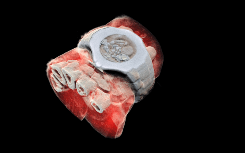
What if, instead of a black and white X-ray picture, a doctor of a cancer patient had access to colour images identifying the tissues being scanned? This colour X-ray imaging technique could produce clearer and more accurate pictures and help doctors give their patients more accurate diagnoses.
This is now a reality, thanks to a New-Zealand company that scanned, for the first time, a human body using a breakthrough colour medical scanner based on the Medipix3 technology developed at CERN. Father and son scientists Professors Phil and Anthony Butler from Canterbury and Otago Universities spent a decade building and refining their product.
Medipix is a family of read-out chips for particle imaging and detection. The original concept of Medipix is that it works like a camera, detecting and counting each individual particle hitting the pixels when its electronic shutter is open. This enables high-resolution, high-contrast, very reliable images, making it unique for imaging applications in particular in the medical field.
Hybrid pixel-detector technology was initially developed to address the needs of particle tracking at the Large Hadron Collider, and successive generations of Medipix chips have demonstrated over 20 years the great potential of the technology outside of high-energy physics.
MARS Bioimaging Ltd, which is commercialising the 3D scanner, is linked to the Universities of Otago and Canterbury. The latter, together with more than 20 research institutes, forms the third generation of the Medipix collaboration. The Medipix3 chip is the most advanced chip available today and Professor Phil Butler recognises that “this technology sets the machine apart diagnostically because its small pixels and accurate energy resolution mean that this new imaging tool is able to get images that no other imaging tool can achieve.”
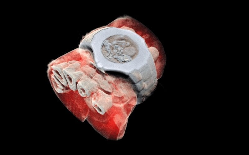
MARS’ solution couples the spectroscopic information generated by the Medipix3 enabled detector with powerful algorithms to generate 3D images. The colours represent different energy levels of the X-ray photons as recorded by the detector and hence identifying different components of body parts such as fat, water, calcium, and disease markers.
So far, researchers have been using a small version of the MARS scanner to study cancer, bone and joint health, and vascular diseases that cause heart attacks and strokes. “In all of these studies, promising early results suggest that when spectral imaging is routinely used in clinics it will enable more accurate diagnosis and personalisation of treatment,” Professor Anthony Butler says.
CERN’s Knowledge Transfer group has a long-standing expertise in transferring CERN technologies, in particular for medical applications. In the case of the 3D scanner, a licence agreement has been established between CERN, on behalf of the Medipix3 collaboration and MARS Bioimaging Ltd. As Aurélie Pezous, CERN Knowledge Transfer Officer states: “It is always satisfying to see our work leveraging benefits for patients around the world. Real-life applications such as this one fuels our efforts to reach even further.”
In the coming months, orthopaedic and rheumatology patients in New Zealand will be scanned by the revolutionary MARS scanner in a clinical trial that is a world first, paving the way for a potentially routine use of this new generation equipment.
The Latest on: Colour medical scanner
[google_news title=”” keyword=”colour medical scanner” num_posts=”10″ blurb_length=”0″ show_thumb=”left”]
via Google News
The Latest on: Colour medical scanner
- Sheriff: Many reasons for mounting jail deathson April 24, 2024 at 1:30 am
Jail personnel no longer just have to worry about suicide while watching inmates. Drugs, medical issues and mental health also have led to increased jail deaths. A record four inmate deaths took place ...
- ‘Comprehensive and life-saving’: State’s attorney introduces new plan aiming to prevent gun violence in Waukegan, North Chicago and Zionon April 23, 2024 at 4:40 pm
The Lake County state's attorney, joined by local and state officials, announced a new "violence reduction plan" Tuesday afternoon that aims to prevent gun violence in the Waukegan, North Chicago and ...
- 3D Scanning Market Mastering Consumer Behavior Insights into Observational Revelationson April 21, 2024 at 9:35 pm
The 3D scanning is a process in which three-dimensional attributes of an object are captured along with information such as colour and texture. The technology saves time, cost and efforts during the ...
- The Best Scanners for 2024on April 18, 2024 at 5:00 pm
Fujitsu ScanSnap iX1300 Compact Wireless Color Scanner — $259.99 (List Price $348) Epson Workforce ES-580W Wireless Color Document Scanner — $349.99 (List Price $429.99) *Deals are selected by ...
- 6 ways to celebrate emergency medical dispatcherson April 16, 2024 at 1:26 pm
Consider these gifts and tips for showing your appreciation during National Public Safety Telecommunicators Week ...
- The Best Portable Scanners for 2024on March 30, 2024 at 5:01 pm
Fujitsu ScanSnap iX1300 Compact Wireless Color Scanner — $259.99 (List Price $348) Epson Workforce ES-580W Wireless Color Document Scanner — $349.99 (List Price $429.99) *Deals are selected by ...
- Free yourself from clutter with the best document scanners in 2024on March 11, 2024 at 11:37 am
Using one of the best document or photo scanners, it's easy and quick to create digital versions of important papers or photos and keep them stored securely in your computer or the cloud. And once ...
- Epson WorkForce DS-770 Color Document Scanneron January 28, 2022 at 1:18 am
PCMag PCMag.com and PC Magazine are among the federally registered trademarks of Ziff Davis, LLC and may not be used by third parties without explicit permission. The ...
- book scanneron April 28, 2016 at 5:00 pm
[Daniel Reetz] spent six years working as a Disney engineer during the day and on his book scanner, the archivist ... one needs a light that doesn’t lose any color information.
via Bing News


