


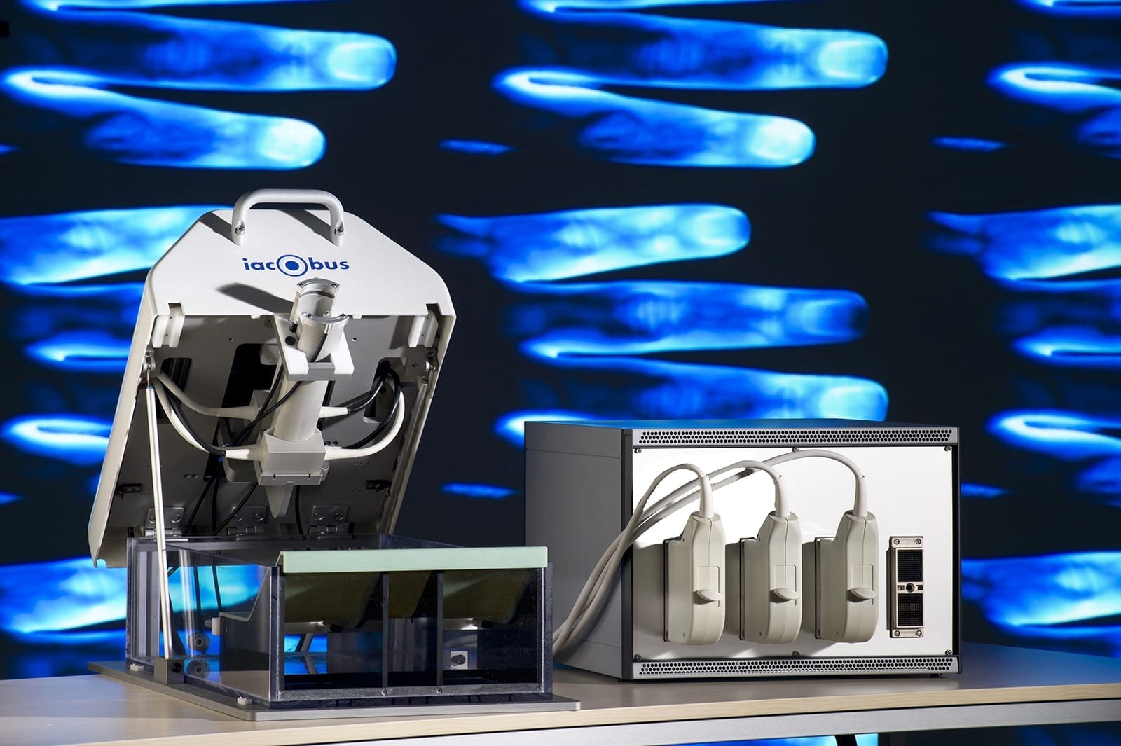
Fraunhofer IBMT/Bernd Müller
Joint inflammation (arthritis) is a common problem in medical practice and can be due to a variety of causes. Many types of inflammatory disorders affecting the joints belong to the diverse group of rheumatic diseases. The most common ones are rheumatoid arthritis and osteoarthritis which frequently affect the joints of the hands. These joint diseases are chronic in nature and cannot be cured yet.
However, an early diagnosis and thus early medical treatment tremendously improves long-term outcome. That is why experts working on the EC-funded project IACOBUS led by the Fraunhofer Institute for Biomedical Engineering IBMT are developing a finger scanner which in the future will allow arthritis of the hands to be diagnosed at a very early stage. The team will be presenting a prototype of the new technology at the MEDICA trade fair in Düsseldorf from November 16 – 19 (Hall 10, Booth G05).
Human joints are incredibly high-tech. Layers of smooth cartilage facilitate swift motion of the bones without friction and the inner lining of the joint capsule, the so-called synovial membrane, shrouds the joint in a casing that constantly produces its own lubricant. For individuals suffering from chronic arthritis, however, this process is severely disturbed by an inflammation of the synovial membrane – which is most severe and destructive in cases of rheumatoid arthritis. Over time, inflammation of the synovium results in damage to the cartilage and even the bones of the joints thus causing severe pain and stiffening of the joints.
Chronic arthritis such as rheumatoid arthritis has no cure as yet, but when caught at an early stage it can be held in check successfully using medication. However, early detection of arthritis requires suitable imaging techniques. Conventional X-ray imaging will only detect typical features of arthritis at a fairly advanced stage. By contrast, use of Doppler ultrasound is more likely to detect arthritis at an earlier stage by visualizing changes in local blood flow. Increased blood flow in the inflamed and thickened synovial membrane is a typical sign of the condition, caused not only by widening of existing blood vessels but also by formation of new blood vessels as a result of the inflammatory process. However, since formation of blood vessels starts off very small with a correspondingly low blood flow, its detection by Doppler ultrasound at an early stage still remains challenging. Magnetic resonance imaging (MRI) can detect arthritic changes of cartilage and bone earlier than X-ray but is significantly more expensive than X-ray and Doppler ultrasound.
Scanner searches joints for sites of inflammation
To improve the early detection of different types of arthritis, a European consortium composed of several research institutions and companies led by the Fraunhofer Institute for Biomedical Engineering IBMT in Saarland, Germany is currently developing an alternative diagnostic technique combining ultrasound technology with novel detection methods. Specifically, this involves the use of a 3-D finger scanner that searches joints for sites of inflammation as well as other pathological changes. “One of the advantages of this method is that it enables us to detect the condition while it is still in its early stages, since many forms of arthritis affect the fingers first”, says Dr. Marc Fournelle, IACOBUS project manager at Fraunhofer IBMT.
Read more: Detecting arthritis with light

The Latest on: Detecting arthritis
[google_news title=”” keyword=”Detecting arthritis” num_posts=”10″ blurb_length=”0″ show_thumb=”left”]
via Google News
The Latest on: Detecting arthritis
- New Brain Circuit Identified in Mice That Controls Body’s Inflammatory Reactionson May 1, 2024 at 4:01 pm
Future research could identify drugs that can target this newfound brain circuit to help treat a vast range of immune disorders and diseases.
- Scientists identify new brain circuit in mice that controls body's inflammatory reactionson May 1, 2024 at 11:50 am
The brain can direct the immune system to an unexpected degree, capable of detecting, ramping up and tamping down inflammation, shows a new study in mice.
- Patients with rheumatoid arthritis have unique and complex autoantibody patterns, study revealson April 30, 2024 at 9:00 pm
Rheumatoid arthritis is a chronic autoimmune disease that primarily ... Wrongly produced antibodies are then detected and removed. Bondt suspects that glycans could help ACPAs trick the control system ...
- Patients with rheumatoid arthritis have unique and complex autoantibody patternson April 30, 2024 at 5:00 pm
Patients with rheumatoid arthritis (RA) all have a unique and diverse set of antibodies that are involved in the development of the disease. Researchers at Utrecht University unveiled the complexity ...
- Rheumatoid Arthritis Patients Have Unique Autoantibody Patternson April 30, 2024 at 5:00 pm
Rheumatoid arthritis is a chronic autoimmune disease that primarily ... Wrongly produced antibodies are then detected and removed. Bondt suspects that glycans could help ACPAs trick the control system ...
- Blood test detects signs of osteoarthritis 8 years before X-rayon April 29, 2024 at 11:34 am
Researchers have created a new blood test that could lead to earlier intervention for those with osteoarthritis.
- AI-powered test predicts knee osteoarthritis 8 years before X-rayon April 27, 2024 at 10:33 am
A new biomarker test successfully predicted the early onset of knee osteoarthritis years ahead of noticeable signs.
- If detected early, osteoarthritis can be 'arrested' to restore joint healthon April 27, 2024 at 9:26 am
A team of researchers in the US predicted knee osteoarthritis via a blood test at least eight years before tell-tale signs of the disease appeared on.health. healthcare. fitness. wellness. well-being.
- Blood test finds knee osteoarthritis up to 8 years before it appears on X-rayson April 26, 2024 at 3:22 pm
Detecting knee osteoarthritis with a blood test up to eight years before it appears on X-rays -- as researchers have done -- could lead to therapies that delay disease progression and restore joint ...
- Blood test may be able to detect osteoarthritis 8 years earlier than X-rayon April 26, 2024 at 12:00 pm
A blood test could help predict knee osteoarthritis at least eight years before the signs of the disease show up on X-rays, a new study indicates.
via Bing News