


New attachment turns a smartphone into a microscope that can image and size DNA molecules 50,000 times thinner than a human hair
If you thought scanning one of those strange, square QR codes with your phone was somewhat advanced, hold on to your seat. Researchers at the University of California, Los Angeles (UCLA) have recently developed a device that can turn any smartphone into a DNA-scanning fluorescent microscope.
“A single DNA molecule, once stretched, is about two nanometers in width,” said Aydogan Ozcan, HHMI Chancellor Professor, UCLA. “For perspective, that makes DNA about 50,000 times thinner than a human hair. Currently, imaging single DNA molecules requires bulky, expensive optical microscopy tools, which are mostly confined to advanced laboratory settings. In comparison, the components for my device are significantly less expensive.”
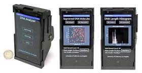
Enter Ozcan’s smartphone attachment — an external lens, thin-film interference filter, miniature dovetail stage mount for making fine alignments, and a laser diode, all enclosed in a small, 3D-printed case and integrated to act just like a fluorescence microscope.
Although other smart-phone-turned-microscopes can image larger scale objects such as cells, Ozcan’s group’s latest mobile-phone optical attachment is the first to image and size the slim strand of a single DNA molecule.
The device is intended for use in remote laboratory settings to diagnose various types of cancers and nervous system disorders, such as Alzheimer’s, as well as detect drug resistance in infectious diseases. To use the camera it is necessary to first isolate and label the desired DNA with fluorescent tags. Ozcan says such laboratory procedures are possible even in remote locations and resource-limited settings.
To scan the DNA, the group developed a computational interface and Windows smart application running on the same smart phone. The scanned information is then sent to a remote server in Ozcan’s laboratory, which measures the length of the DNA molecules. Assuming you have a reliable data connection, the entire data processing takes less than 10 seconds.
Read more: A Phone with the Ultimate Macro Feature
The Latest on: DNA-scanning fluorescent microscope
[google_news title=”” keyword=”DNA-scanning fluorescent microscope” num_posts=”10″ blurb_length=”0″ show_thumb=”left”]
via Google News
The Latest on: DNA-scanning fluorescent microscope
- Rapid nanoplasmonic-enhanced detection of SARS-CoV-2 and variants on DNA aptamer metasurfaceson April 27, 2024 at 5:00 pm
Flowchart for genetic algorithm and computational screening of periodic gold nanostructures for maximizing the Raman cross-section of the metasurfaces, Scanning emission microscopy images of the e ...
- Advanced microscopy technique offers a new look inside cellson April 10, 2024 at 5:00 pm
Researchers face a similar dilemma when imaging cells under a microscope ... imaging probes consisting of a single strand of DNA and a fluorescent dye. The antibody guides the probe to its ...
- Scanning Tunneling Microscopy STMon March 20, 2024 at 5:00 pm
The leading scientific social networking website and producer of educational virtual events and webinars.
- DNA Sequencing Technologieson November 11, 2023 at 5:17 am
In 1986, a company named Applied Biosystems began to manufacture automated DNA sequencing machines ... These include flow cytometry, scanning microscopes, mass spectrometry, and hybridization ...
- Light Microscopy Facilityon November 12, 2021 at 3:34 pm
The equipment includes two confocal microscopes for imaging fixed and live samples, and four slide scanners for brightfield and fluorescent ... locally cause DNA damage and observe the effects of this ...
- DVD Optics Power This Scanning Laser Microscopeon February 1, 2021 at 5:16 pm
The basic scheme here is known as confocal laser scanning fluorescence microscopy, where a laser at one wavelength excites fluorescent tags bound to structures in a sample. Light emitted by the ...
- Fluorescent and Confocal Microscopyon December 18, 2020 at 3:14 am
The Pathology Core laboratory is equipped with a Nikon E-1000 upright fluorescent microscope equipped with 4x, 10x, 20x and 40x dry objectives and 40x and 60x oil immersion objectives. The microscope ...
- Fluorescence Microscopyon March 29, 2019 at 5:00 pm
Some of biology’s most visually striking images come from fluorescence microscopes. Their brilliant colors on black look like a neon sign from an empty highway. A brand new fluorescence ...
- Fluorescence Optical Microscopy Facilityon February 27, 2019 at 9:04 am
The capabilities of the shared microscopy facility include brightfield, phase contrast, and both 2-D and 3-D fluorescence imaging. The OLYMPUS BX51 research microscope is a union of Olympus infinity ...
- Enhancing the power of super-resolution microscopyon August 21, 2018 at 5:00 pm
Previous fluorescent markers for target proteins have been too large and this has been a key factor slowing down the progression of super-resolution microscopy. Therefore, the team decided to downsize ...
via Bing News

