via phys.org
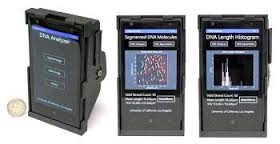
New attachment turns a smartphone into a microscope that can image and size DNA molecules 50,000 times thinner than a human hair
If you thought scanning one of those strange, square QR codes with your phone was somewhat advanced, hold on to your seat. Researchers at the University of California, Los Angeles (UCLA) have recently developed a device that can turn any smartphone into a DNA-scanning fluorescent microscope.
“A single DNA molecule, once stretched, is about two nanometers in width,” said Aydogan Ozcan, HHMI Chancellor Professor, UCLA. “For perspective, that makes DNA about 50,000 times thinner than a human hair. Currently, imaging single DNA molecules requires bulky, expensive optical microscopy tools, which are mostly confined to advanced laboratory settings. In comparison, the components for my device are significantly less expensive.”
Enter Ozcan’s smartphone attachment — an external lens, thin-film interference filter, miniature dovetail stage mount for making fine alignments, and a laser diode, all enclosed in a small, 3D-printed case and integrated to act just like a fluorescence microscope.
Although other smart-phone-turned-microscopes can image larger scale objects such as cells, Ozcan’s group’s latest mobile-phone optical attachment is the first to image and size the slim strand of a single DNA molecule.
The device is intended for use in remote laboratory settings to diagnose various types of cancers and nervous system disorders, such as Alzheimer’s, as well as detect drug resistance in infectious diseases. To use the camera it is necessary to first isolate and label the desired DNA with fluorescent tags. Ozcan says such laboratory procedures are possible even in remote locations and resource-limited settings.
To scan the DNA, the group developed a computational interface and Windows smart application running on the same smart phone. The scanned information is then sent to a remote server in Ozcan’s laboratory, which measures the length of the DNA molecules. Assuming you have a reliable data connection, the entire data processing takes less than 10 seconds.
Read more: A Phone with the Ultimate Macro Feature
The Latest on: DNA-scanning fluorescent microscope
[google_news title=”” keyword=”DNA-scanning fluorescent microscope” num_posts=”10″ blurb_length=”0″ show_thumb=”left”]
via Google News
The Latest on: DNA-scanning fluorescent microscope
- Testing how well biomarkers work: New fluorescence microscopy method can improve resolution down to the Ångström scaleon April 24, 2024 at 9:49 am
LMU researchers have developed a method to determine how reliably target proteins can be labeled using super-resolution fluorescence microscopy.
- Research combines DNA origami and photolithography to move one step closer to molecular computerson April 24, 2024 at 8:49 am
Molecular computer components could represent a new IT revolution and help us create cheaper, faster, smaller, and more powerful computers. Yet researchers struggle to find ways to assemble them more ...
- An ultracompact multimode meta-microscopeon March 29, 2024 at 11:20 am
In conclusion, the researchers propose and demonstrate a miniaturized multimode meta-microscope based on guided-wave illumination. Three imaging modes are realized within a centimeter-scale ...
- Confocal MEMS unit for fluorescence imagingon June 6, 2023 at 1:35 am
The MEMS confocal unit can be connected to an inverted microscope to allow ... sectioning images utilizing a spot scanning device and pinholes. Only fluorescence emitted from the focus plane ...
- DVD Optics Power This Scanning Laser Microscopeon February 1, 2021 at 5:16 pm
The basic scheme here is known as confocal laser scanning fluorescence microscopy, where a laser at one wavelength excites fluorescent tags bound to structures in a sample. Light emitted by the ...
- confocal microscopyon February 1, 2021 at 4:00 pm
The basic scheme here is known as confocal laser scanning fluorescence microscopy, where a laser at one wavelength excites fluorescent tags bound to structures in a sample. Light emitted by the ...
- Enhancing the power of super-resolution microscopyon August 21, 2018 at 5:00 pm
Previous fluorescent markers for target proteins have been too large and this has been a key factor slowing down the progression of super-resolution microscopy. Therefore, the team decided to downsize ...
- Scanning Electron Microscopeon October 13, 2017 at 5:10 pm
SEM stands for scanning electron microscope. The SEM is a microscope that uses electrons instead of light to form an image. Since their development in the early 1950's, scanning electron microscopes ...
- Practical considerations for fluorescence microscopyon June 28, 2016 at 5:59 am
In this presentation, we will go over some of the reasons you would want to consider using fluorescence imaging and give a brief introduction to using fluorescent probes for cell structure and ...
- Equipment Liston November 26, 2015 at 6:26 am
OD range 200 to 1,000 and Fluorescence Excitation 230-850 and Emmission 280 to 850. DNA/RNA NanoQuant plate measurements. For the measurement of particle and molecule size from below a nanometer to ...
via Bing News


