
Source: College of Engineering A noninvasive skin patch is one way the researchers’ ultrasound-based method could allow for easy imaging of the brain.
One day, scopes may no longer need to be inserted into the body, such as down the throat or under the skin, to reach the stomach, brain, or any other organs for examination.
Maysam Chamanzar, assistant professor of electrical and computer engineering, and Matteo Giuseppe Scopelliti, an ECE Ph.D. student, have introduced a novel technique that uses ultrasound to noninvasively take optical images through a turbid medium such as biological tissue to image body’s organs. This new method has the potential to eliminate the need for invasive visual exams using endoscopic cameras.
Endoscopic imaging, or using cameras inserted directly inside the body’s organs to investigate symptoms, is an invasive procedure used to examine and diagnose symptoms of deep tissue disease. Endoscopic imagers, or cameras on the end of catheter tubes or wires, are usually implanted through a medical procedure or surgery in order to reach the body’s deep tissues. Chamanzar’s new technique provides a completely non-surgical and noninvasive alternative.
The lab’s paper, published in Light: Science and Applications, a journal published by Springer Nature, shows that they can use ultrasound to create a virtual “lens” within the body, rather than implanting a physical lens. By using ultrasonic wave patterns, the researchers can effectively “focus” light within the tissue, which allows them to take images never before accessible through noninvasive means.
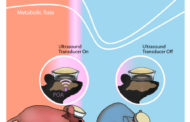
Biological tissue is able to block most light, especially light in the visible range of the optical spectrum. Therefore, current optical imaging methods cannot use light to access deep tissue from the surface. Chamanzar’s lab, however, has used noninvasive ultrasound to induce more transparency to enable more penetration of light through turbid media, such as biological tissue.
“Being able to relay images from organs, such as the brain, without the need to insert physical optical components will provide an important alternative to implanting invasive endoscopes in the body,” says Chamanzar. “We used ultrasound waves to sculpt a virtual optical relay lens within a given target medium, which for example, can be biological tissue. Therefore, the tissue is turned into a lens that helps us capture and relay the images of deeper structures.”
Being able to relay images from organs such as the brain without the need to insert physical optical components will provide an important alternative to implanting invasive endoscopes in the body.
Maysam Chamanzar, Assistant Professor, Electrical and Computer Engineering
“This method can revolutionize the field of biomedical imaging,” says Chamanzar.
Ultrasound waves are able to compress and rarefy, or thin, whatever medium they are flowing through. In compressed regions, light travels more slowly compared to rarefied regions. In this paper, the team shows that this compression and rarefication effect can be used to sculpt a virtual lens in the target medium for optical imaging. This virtual lens can be moved around without disturbing the medium simply by reconfiguring the ultrasound waves from outside. This enables imaging different target regions, all noninvasively.
The published method is a platform technology that can be applied in many different applications. In the future, it can be implemented in the form of a handheld device or wearable surface patch, depending on the organ being imaged. By placing the device or patch on the skin, the clinician would be able to easily receive optical information from within the tissue to create images of what’s inside without endoscopy’s many discomforts and side effects.
The closest current applications for this technology would be endoscopic imaging of brain tissue or imaging under the skin, but this technique can also be used in other parts of the body for imaging. Beyond biomedical applications, this technique can be used for optical imaging in machine vision, metrology, and other industrial applications to enable non-destructive and steerable imaging of objects and structures at the micron scale.
The researchers showed that the properties of the virtual “lens” can be tuned by changing the parameters of the ultrasonic waves, allowing users to “focus” images taken using the method at different depths through the medium. While the LSA paper is focused on the method’s efficacy for closer-to-the-surface applications, the team has yet to find the limit to how deep within the body’s tissue this ultrasonically-assisted optical imaging method can reach.
“What distinguishes our work from conventional acousto-optic methods is that we are using the target medium itself, which can be biological tissue, to affect light as it propagates through the medium,” explains Chamanzar. “This in situ interaction provides opportunities to counterbalance the non-idealities that disturb the trajectory of light.”
This technique has many potential clinical applications, such as diagnosing skin disease, monitoring brain activity, and diagnosis and photodynamic therapy for identifying and targeting malignant tumors.
In addition to the direct implications this research has on clinical medicine, it will also have indirect clinical applications. By using this acousto-optic technology to view mouse models of brain disorders in action and selectively stimulate different neural pathways, researchers would be able to study the mechanisms involved in disease conditions such as Parkinson’s, informing the design of next generation clinical therapeutic interventions to treat these diseases in humans.
“Turbid media have always been considered obstacles for optical imaging,” says Scopelliti. “But we have shown that such media can be converted to allies to help light reach the desired target. When we activate ultrasound with the proper pattern, the turbid medium becomes immediately transparent. It is exciting to think about the potential impact of this method on a wide range of fields, from biomedical applications to computer vision.”
The researchers project that this new imaging technology could be applied in biomedical and clinical contexts within the next five years.
Learn and see more: Breakthrough in optical endoscopy using ultrasound
The Latest on: Biomedical imaging
[google_news title=”” keyword=”biomedical imaging” num_posts=”10″ blurb_length=”0″ show_thumb=”left”]
via Google News
The Latest on: Biomedical imaging
- New imaging technique provides a much closer look at fibril assemblieson April 26, 2024 at 10:47 pm
A new imaging technique developed by engineers at Washington University in St. Louis can give scientists a much closer look at fibril assemblies, stacks of peptides like amyloid beta, most notably ...
- Library Faculty to Help Researchers Access Massive Biomedical Databaseon April 25, 2024 at 11:28 am
UM Libraries received the $40,000 All of Us Academic Libraries Program award. Library faculty Savannah Kelly and Shelby Watson will offer workshops for both beginning and veteran researchers on how to ...
- Perinatal substance use may shape how strongly mothers feel toward infantson April 25, 2024 at 10:22 am
Substance use during pregnancy and postpartum may impact areas of the brain associated with what psychologists and neuroscientists call "affective empathy," or the emotional response experienced as a ...
- Biomedical Engineer Salaryon April 24, 2024 at 5:00 pm
How Much Does a Biomedical Engineer Make? Biomedical Engineers made a median salary of $99,550 in 2022. The best-paid 25% made $129,230 that year, while the lowest-paid 25% made $78,500.
- Imaging technique shows new details of peptide structureson April 24, 2024 at 11:19 am
A new imaging technique developed by engineers at Washington University in St. Louis can give scientists a much closer look at fibril assemblies—stacks of peptides that include amyloid beta, most ...
- Biomedical Applications of Nanodiamonds in Imaging and Therapyon April 19, 2024 at 4:59 pm
In this review an attempt is made to capture the scope, spirit and recent developments in the field of nanodiamonds for biomedical applications ... optimizing them for imaging and sensing ...
- Researchers review use of MRI to identify brain cancer biomarkerson April 11, 2024 at 12:15 pm
Researchers from the School of Biomedical Engineering & Imaging Sciences (BMEIS) have published a systematic review in Neuro-Oncology Advances exploring the use of MRI imaging techniques to identify ...
- Biomedical Applications of Nanodiamonds in Imaging and Therapyon April 7, 2024 at 5:00 pm
In addition to the biomedical applications of NDs in the ... the spectroscopic properties for adjusting/optimizing them for imaging and sensing applications are still under investigation.
- Updated with memorial fund information: Biomedical Engineering Chair Joseph Izatt Dieson April 7, 2024 at 5:00 pm
The lab’s expertise in OCT technology has also allowed them to expand their reach beyond the realm of biomedical imaging to other endeavors, such as investigating how OCT could help autonomous robots ...
- Data-driven polarimetric imagingon April 3, 2024 at 5:00 pm
Existing data-driven polarization imaging technologies have gradually found applications in polarization information reconstruction and enhancement, target detection, biomedical imaging ...
via Bing News


