


Researchers at the University of Rochester Medical Center have developed a new imaging technique that could revolutionize how eye health and disease are assessed. The group is first to be able to make out individual cells at the back of the eye that are implicated in vision loss in diseases like glaucoma. They hope their new technique could prevent vision loss via earlier diagnosis and treatment for these diseases.
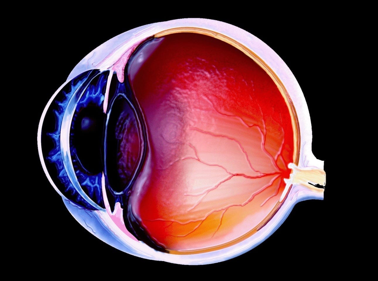
In a study highlighted in the Proceedings of the National Academy of Sciences, Ethan A. Rossi, Ph.D., assistant professor of Ophthalmology at the University of Pittsburgh School of Medicine, describes a new method to non-invasively image the human retina, a layer of cells at the back of the eye that are essential for vision. The group, led by David Williams, Ph.D., Dean for Research in Arts, Sciences, and Engineering and the William G. Allyn Chair for Medical Optics at the University of Rochester, was able to distinguish individual retinal ganglion cells (RGCs), which bear most of the responsibility of relaying visual information to the brain.
There has been a longstanding interest in imaging RGCs because their death causes vision loss in glaucoma, the second leading cause of acquired blindness worldwide. Despite great efforts, no one has successfully captured images of individual human RGCs, in part because they are nearly perfectly transparent.
This new approach might eventually allow us to detect the loss of single ganglion cells. The sooner we can catch the loss, the better our chances of halting disease and preventing vision loss.
Instead of imaging RGCs directly, glaucoma is currently diagnosed by assessing the thickness of the nerve fibers projecting from the RGCs to the brain. However, by the time a change is typically detected in the retinal nerve fiber thickness, a patient may have lost tens of thousands of RGCs or more.
“In principle, this new approach might eventually allow us to detect the loss of single ganglion cells,” said Williams. “The sooner we can catch the loss, the better our chances of halting disease and preventing vision loss.”
Rossi and his colleagues were able to see RGCs by modifying an existing technology – confocal adaptive optics scanning light ophthalmoscopy (AOSLO). They collected multiple images, varying the size and location of the detector they used to gather light scattered out of the retina for each image, and then combined those images. The technique, called multi-offset detection, was performed at the University of Rochester Medical Center in animals as well as volunteers with normal vision and patients with age-related macular degeneration.
While RGCs were the main focus of Rossi’s investigations, they are just one type of cell that can be imaged using this new technique. In age-related macular degeneration, cone photoreceptors that detect color and are important for central vision are the first to die. AOSLO has been used to image cones before, but these cells were difficult to see in areas near Drusen, fatty deposits that are the most common early sign of the disease. Using their multi-offset technique in age-related macular degeneration patients, Rossi was able to assess the health of cones near Drusen and in areas where the retina had been damaged.
“This technique offers the opportunity to evaluate many retinal features that have previously remained inaccessible to imaging in the living eye,” said Rossi. “Not only RGCs, but potentially other nearly transparent cell classes as well.”
Rossi and his colleagues warn that their study included a small number of volunteers and an even smaller number of age-related macular degeneration patients. More studies will be needed to improve the robustness of the technique before it can be widely used in the clinic. Rossi is now setting up his own laboratory at the University of Pittsburgh and plans to continue working with Williams’ group in studying this technique and its ability to detect changes in retinal cells over the course of retinal diseases.
Learn more: A Closer Look at the Eye: Researchers Develop New Retinal Imaging Technique
[osd_subscribe categories=’eye-disease-detection’ placeholder=’Email Address’ button_text=’Subscribe Now for any new posts on the topic “EYE DISEASE DETECTION”‘]
Receive an email update when we add a new EYE DISEASE DETECTION article.
The Latest on: Eye disease detection
[google_news title=”” keyword=”eye disease detection” num_posts=”10″ blurb_length=”0″ show_thumb=”left”]
via Google News
The Latest on: Eye disease detection
- The Canadian Association of Optometrists Releases a New “GetEyeWise” Digital Campaign to Bring Awareness About Eye Health and Vision Careon May 1, 2024 at 10:46 am
As May marks Vision Health Month, the Canadian Association of Optometrists (CAO) releases a digital national campaign to raise publi ...
- OPTOMED RELEASES FIRST FDA-CLEARED HANDHELD AI FUNDUS CAMERA FOR DETECTION OF MORE THAN MILD DIABETIC RETINOPATHYon May 1, 2024 at 7:00 am
Optomed USA, Inc., a medical technology company, introduces the Optomed Aurora AEYE, a handheld AI fundus camera that provides instant detection of more than mild diabetic retinopathy. With the ...
- Toku bags breakthrough designation for kidney disease eye scan deviceon May 1, 2024 at 4:22 am
The US company says the system works by scanning the retinas for signs of chronic kidney disease in a bid to catch the disease before it progresses.
- Optomed Oyj, AEYE Health say portable device to detect eye issues gets FDA nodon May 1, 2024 at 3:28 am
A portable device that detects eye conditions that can cause sight loss in people with conditions such as diabetes has received clearance from the U.S. health regulator, its developers Optomed Oyj and ...
- Seeing the future: How a New Mexico start-up is testing AI for detecting eye diseaseon April 30, 2024 at 4:07 pm
Twenty years from now, most diagnostics within primary care will be done using the aid of artificial intelligence, at least, that’s the goal of a New ...
- America’s Infectious-Disease Barometer Is Offon April 30, 2024 at 12:44 pm
Somehow, the U.S. is both over- and under-reacting to bird flu and other pressing infectious threats. The ongoing outbreak of H5N1 avian flu virus looks a lot like a public-health problem that the ...
- AI-Powered Solutions for Personalized Therapeutic Interventions in Dry Eye Diseaseon April 24, 2024 at 12:44 pm
According to a study published in Big Data Mining and Analytics, researchers want to employ artificial intelligence (AI) to help in the early detection and diagnosis of Dry Eye Disease (DED).
- Researchers aim to use AI for early screening and prognosis of Dry Eye Diseaseon April 23, 2024 at 1:19 pm
Dry Eye Disease (DED) is one of the more common eye diseases, affecting up to 30% of the world's population. This disease can affect many different types of people and can wind up being a great ...
- Artificial Intelligence Helps Detect Dry Eye Diseaseon April 23, 2024 at 11:07 am
AI's ongoing learning process acts as a catalyst, driving research forward by contributing to predictive models for dry eye disease.
via Bing News