


Purdue University researchers are developing a novel biomedical imaging system that combines optical and ultrasound technology to improve diagnosis of life-threatening diseases.
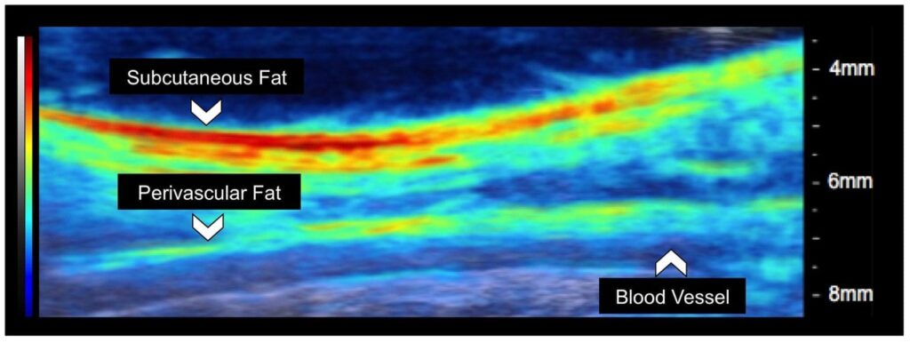
Photoacoustic tomography is a noninvasive technique that works by converting absorbed optical energy into acoustic signal. Pulsed light is sent into body tissue, creating a small increase in temperature that causes the tissue to expand and create an acoustic response that can be detected by an ultrasound transducer. The ultrasound data is used to visualize the tissue.
“The nice thing about photoacoustic tomography is the compositional information,” said Craig Goergen, an assistant professor in Purdue’s Weldon School of Biomedical Engineering. “It provides information about where blood and lipid are located, along with other essential information.”
The ultimate goal is to enhance the clinical care of patients.
The results of a study describing an adjustable photoacoustic probe with improved light delivery and image quality were published Tuesday (Aug. 28) in the journal Photoacoustics.
The system provides real-time compositional information of body tissue without the need for contrast agents and with better depth penetration compared with conventional optical techniques.
Photoacoustic tomography can be used to detect or monitor a myriad of diseases, including cardiovascular disease, diabetes, and cancer. Those are diseases that the Centers for Disease Control and Prevention lists as among the most common, costly, and preventable of all health problems. Heart disease and cancer each account for one in every four deaths a year in the United States, and more than 30 million Americans, or more than 9 percent of the population, have diabetes. The cost of those three diseases a year in the United States is more than $718 billion a year, according to the CDC.
“That means there will be a great need for medical imaging. Trying to diagnose these diseases at an earlier time can lead to improved patient care,” Goergen said. “We are in the process now of trying to use this enhanced imaging approach to a variety of different applications to see what it can be used for.”
Among other potential uses for photoacoustic tomography is the mapping of lipid deposition within an arterial wall that can cause other health problems, measuring cardiac tissue damage and tumor biopsies. Using photoacoustic tomography for intraoperative tumor biopsies could help surgeons make sure they remove all the cancer from a patient, Goergen said.
One of the challenges of photoacoustic tomography is improving the penetration depth and signal-to-noise ratio past optical absorbers. The researchers believe creating optical manipulation techniques to maximize photon density could provide a solution. As a result, they have created a motorized photoacoustic holder that allows users to easily maneuver the aim of the device and tune the depth where light is focused, improving the light penetration depth and signal-to-noise ratio.
A video about the acoustic tomography is available at https://bit.ly/2yJddb0. A complete list of co-authors is available in the abstract. The research has been funded by the National Institutes of Health.
The Purdue researchers are interested in talking with other companies about other possible uses for photoacoustic tomography. The researchers have a patent pending for the technology with the help of the Purdue Office of Technology Commercialization.
Learn more: Purdue researchers developing novel biomedical imaging system combining optical, ultrasound technology

The Latest on: Biomedical imaging
[google_news title=”” keyword=”biomedical imaging” num_posts=”10″ blurb_length=”0″ show_thumb=”left”]
via Google News
The Latest on: Biomedical imaging
- Aroostook breast cancer detection innovator vies for statewide business awardon May 11, 2024 at 5:45 am
Kendra Batchelder, co-founder and CEO of Waved Medical LLC in Presque Isle, is one of two County entrepreneurs set to compete for $25,000 in the state Top Gun Showcase Pitch Event on May 16 in ...
- Biomedical Engineer Salaryon May 9, 2024 at 4:59 pm
How Much Does a Biomedical Engineer Make? Biomedical Engineers made a median salary of $99,550 in 2022. The best-paid 25% made $129,230 that year, while the lowest-paid 25% made $78,500.
- UB awarded $1.77 million grant to create toolset for oxygen metabolism mappingon May 9, 2024 at 4:59 pm
assistant professor in the Department of Biomedical Engineering, the team will use the award to develop new technology that, when paired with common MRI scanners, provides accurate and reliable data ...
- ‘Universally loved’: Joseph Izatt remembered for pioneering biomedical research, inspirational legacyon May 8, 2024 at 8:17 pm
Izatt first joined the BME department at Duke in 2001. Throughout his 23-year tenure, Izatt received the 2008 Capers and Marion McDonald Award for Excellence and Advising from the Pratt School of ...
- New Harvard Technology Paves the Way for Advanced Machine Visionon May 6, 2024 at 3:21 pm
Scientists have developed a compact, single-shot, and complete polarization imaging system using metasurfaces. Think of all the information we get based on how an object interacts with wavelengths of ...
- Said@Duke: Dr. Daniel Saurborn on Combining Studies In Biomedical Engineering, English and Medicineon May 6, 2024 at 11:21 am
Duke alumnus Dr. Daniel Saurborn recenctly spoke at a Duke Humanities in Medicine (HuMed) Celebration. Saurborn is a physician-entrepreneur, biomedical engineering and English double major graduate ...
- Real-time MRI reveals the movement dynamics of stutteringon May 3, 2024 at 6:28 am
Researchers at the University Medical Center Göttingen (UMG) and the Max Planck Institute for Multidisciplinary Sciences (MPI-NAT) have succeeded in visualizing the movement patterns of the internal ...
- Unveiling a polarized world -- in a single shoton May 1, 2024 at 5:00 pm
the imaging system could unlock the vast potential of polarization imaging for a range of existing and new applications, including biomedical imaging, augmented and virtual reality systems and smart ...
- Imaging Technique Shows New Details of Peptide Structureson April 29, 2024 at 8:51 am
A new imaging technique developed by engineers at Washington University in St. Louis can give scientists a much closer look at fibril assemblies, stacks of peptides like amyloid beta, most notably ...
- Duke Biomedical Engineering chairman and OCT pioneer Joseph Izatt passes awayon April 29, 2024 at 6:56 am
Joseph Izatt was a skilled researcher and inventor who played a foundational role in the development of optical coherence tomography.
via Bing News

