


Optical microscopy experts at Colorado State University are once again pushing the envelope of biological imaging.
Jeffrey Field, a research scientist in electrical engineering and director of CSU’s Microscope Imaging Network, has designed and built a fluorescence-detection microscope that combines three-dimensional and high-resolution image processing that’s also faster than comparable techniques.
The work, with co-authorship by Randy Bartels, professor of electrical and computer engineering, and former postdoctoral researcher David Winters, has been published in Optica, the journal of the Optical Society of America. They named their new microscope CHIRPT: Coherent Holographic Image Reconstruction by Phase Transfer.
Imaging tradeoffs
Field and other optics scientists work in a world of tradeoffs. For example: an advanced deep-tissue imaging technique called multiphoton fluorescence microscopy employs a short, bright laser pulse focused tight to one spot, and the fluorescence intensity from that one spot is recorded. Then, the laser moves to the next spot, then the next, to build up high-resolution 3D images. The technique offers subcellular detail, but it’s relatively slow because it illuminates only one tiny spot at a time.
Other techniques, like spinning disk confocal microscopy, are faster because they shine light on multiple spots, not just one, and they scan simultaneously over a larger area. But unlike multiphoton, these techniques require collecting an image with a camera. As a result, fluorescent light emitted from the specimen is blurred on the camera, leading to loss in resolution, and with it, subcellular detail.
Call them greedy, but Field and colleagues want it all.
Breaking established boundaries
Their goal is working around each of these limitations – speed, resolution, size of field – to break through established boundaries in light microscopy.
Field and Bartels’ new microscope builds upon a previously published technique, and permits digital re-focus of fluorescent light. It illuminates not one point, but multiple points by harnessing delocalized illumination spread over a large area. The physical principles they are using are similar to holography, in which scattered light is used to build a 3-D image.
Using a large illumination field, followed by back-end signal processing, the microscope can define distinct light modulation patterns of many points within the field of view. It builds up a 3-D image by combining the signals from all those distinct patterns.
“The idea is that you have a fluorophore at any point in the specimen, and the temporal structure of its fluorescence will be distinguishable from all others,” Field said. “So you can have this huge array of fluorophores, and just with this single-pixel detector, you can tell where every one of them is in that 2D field.”
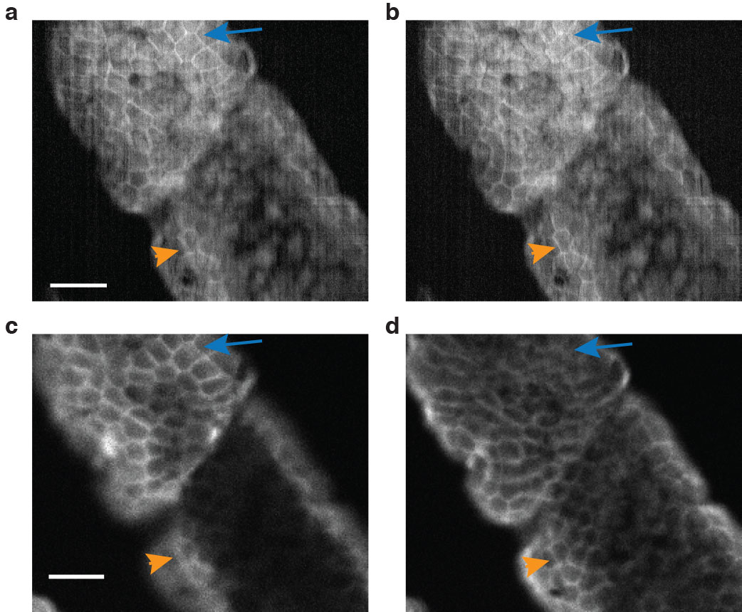
CHIRPT and confocal imaging of fluorescently labelled mouse intestine slices. (a) and (b) are digitally refocused CHIRPT images. (c) and (d) are conventional confocal images. In all four, features in focus are denoted by arrows.
3D, deep-tissue images
So what does this new technique allow? Deep-tissue images in three dimensions, with better depth of field than comparable techniques. Depth of field, like in photography, means background images are in sharp focus along with the main image. And the CSU researchers can work at 600 frames per second, which is many times faster than established techniques.
With their new microscope, images can also be post-processed to remove aberrations that obscure the object of interest. It’s akin to being able to focus a picture after it’s been taken.
The CHIRPT microscope could allow biomedical researchers to produce sharp, 3-D images of cells or tissue over a much larger volume than conventional fluorescence microscopy methods allow. It could lead to things like imaging multicellular processes in real time that, with a conventional light microscope, could only be seen one cell at a time.

Learn more: Fluorescent holography: Upending the world of biological imaging
The Latest on: Fluorescent holography
[google_news title=”” keyword=”Fluorescent holography” num_posts=”10″ blurb_length=”0″ show_thumb=”left”]
via Google News
The Latest on: Fluorescent holography
- Art and Designon May 9, 2024 at 1:35 am
The immersive show features fragile dresses inside airtight vitrines, overcoats growing grass, pat-’n-sniff walls and a hologram ... Tao Siqi’s fluorescent-colored paintings, inspired by ...
- Displays articles from across Nature Portfolioon May 7, 2024 at 5:00 pm
Numerous optical technologies are used in displays, including organic light-emitting diodes and inorganic light-emitting diodes, liquid crystals and fluorescent materials. Important considerations ...
- 29 Mother's Day Gifts That'll Be Sure to Make You the Favorite Childon May 5, 2024 at 5:00 pm
There are lots of unique colors to choose from - my favorite is the Blue Agave, which is a holographic blueish purple ... of blue light from digital devices as well as artificial fluorescent and LED ...
- ultrasonic holographyon April 17, 2024 at 5:00 pm
What distinguishes ultrasonic holography from light-based xolography is that the latter uses photon interference between two light sources in order to rapidly 3D print an object within the print ...
- Researchers made a holographic display from a plain old iPhone 14 screenon April 13, 2024 at 8:22 am
For the most part, though, holographic tech has always seemed a bit outside our wheelhouse, with the tech needed to create realistic holographic displays being exceptionally expensive. Now ...
- New Biden Rule Means Lights Out for Compact Fluorescent Bulbson April 12, 2024 at 2:08 pm
Compact fluorescent light bulbs — once considered corkscrew-shaped emblems of the clean-energy transition — will effectively be phased out under a Biden administration rule finalized Friday ...
- Electron Holography: Unveiling the Nanoscale Worldon April 1, 2024 at 12:09 am
Electron holography is based on the fundamental principles of holography, which involves the recording and reconstruction of wavefronts. In holography, a hologram is created by capturing the ...
- Holographic Cellphones Coming Thanks To AIon January 3, 2023 at 1:35 am
It is easy to envision conference rooms full of computer equipment and scanners, but an MIT student has a method that may do away with all that by using machine learning to simplify hologram ...
- Working at Hologram, Inc.on April 27, 2019 at 2:59 pm
Hologram is a collaborative, remote-first team united by our core values of ownership, transparency, and mindfulness. We trust you to do what’s best for our product, customers, and team members.
via Bing News

