


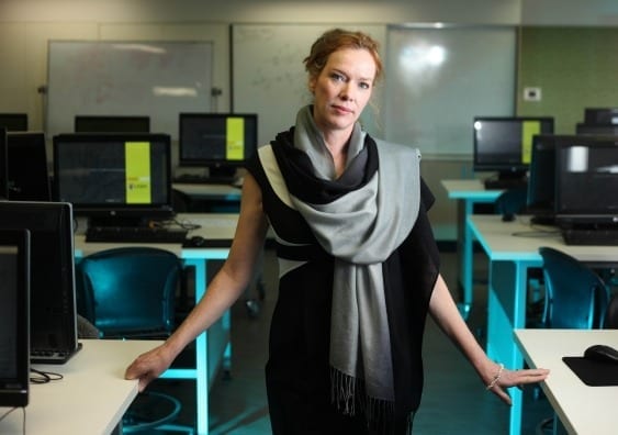
UNSW biomedical engineer Melissa Knothe Tate is using previously top-secret semiconductor technology to zoom through organs of the human body, down to the level of a single cell.
A world-first UNSW collaboration that uses previously top-secret technology to zoom through the human body down to the level of a single cell could be a game-changer for medicine, an international research conference in the United States has been told.
The imaging technology, developed by high-tech German optical and industrial measurement manufacturer Zeiss, was originally developed to scan silicon wafers for defects.
UNSW Professor Melissa Knothe Tate, the Paul Trainor Chair of Biomedical Engineering, is leading the project, which is using semiconductor technology to explore osteoporosis and osteoarthritis.
Using Google algorithms, Professor Knothe Tate – an engineer and expert in cell biology and regenerative medicine – is able to zoom in and out from the scale of the whole joint down to the cellular level “just as you would with Google Maps”, reducing to “a matter of weeks analyses that once took 25 years to complete”.
Her team is also using cutting-edge microtome and MRI technology to examine how movement and weight bearing affects the movement of molecules within joints, exploring the relationship between blood, bone, lymphatics and muscle.
“For the first time we have the ability to go from the whole body down to how the cells are getting their nutrition and how this is all connected,” said Professor Knothe Tate. “This could open the door to as yet unknown new therapies and preventions.”
Professor Knothe Tate is the first to use the system in humans. She has forged a pioneering partnership with the US-based Cleveland Clinic, Brown and Stanford Universities, as well as Zeiss and Google to help crunch terabytes of data gathered from human hip studies.
Similar research is underway at Harvard University and Heidelberg in Germany to map neural pathways and connections in the brains of mice.
Professor Knothe Tate presented several papers on her research into the human hip and osteoarthritis at the peer-reviewed Orthopedic Research Society meeting in Las Vegas.
Numerous studies have explored molecular transport within specific tissues but there has been little research on exchange between different kinds of tissue such as cartilage and bone.
Professor Knothe Tate has already demonstrated a link between molecular transport through blood, muscle and bone, and disease status in osteoarthritic guinea pigs.
Like humans, guinea pigs develop osteoarthritis as they age. The condition is increasingly believed to be the result of a breakdown in cellular communication.
Understanding the molecular signaling and traffic between tissues could unlock a range of treatments, including physical therapies and preventative exercise routines, Professor Knothe Tate said.
Critical to this work has been the development of microscopy that allows seamless imaging of organs and tissues across length scales – centimetres at the whole-joint level down to nanometer-sized molecules – as well as the capacity to sift and analyse huge sets of data.

Professor Knothe Tate likened using the Zeiss technology in the hipbone to Google Maps’ ability to zoom down from an Earth View to Street View.
Read more: ‘Google Maps’ for the body: a biomedical revolution
The Latest on: Biomedical engineering
[google_news title=”” keyword=”Biomedical engineering” num_posts=”10″ blurb_length=”0″ show_thumb=”left”]
via Google News
The Latest on: Biomedical engineering
- Analyzing the Ethics Behind Biomedical Engineeringon May 7, 2024 at 1:12 pm
The movie takes place in an unspecified future where children are ‘made’ through a eugenics-based genome engineering program that removes or limits the risk of diseases and ensures that children ...
- Few tenure-track jobs for engineering PhDson May 6, 2024 at 4:59 pm
Subdisciplines with the lowest placement rates for newly minted PhDs include engineering management, petroleum, biomedical, nuclear engineering, metallurgical and materials, and environmental ...
- LSU's new engineering dean is a Baton Rouge native: See her resume, where she's comign fromon May 6, 2024 at 11:52 am
A Baton Rouge native who currently serves as the director of biomedical engineering at Brown University has been named LSU's new dean of the College of Engineering, the university said ...
- Said@Duke: Dr. Daniel Saurborn on Combining Studies In Biomedical Engineering, English and Medicineon May 6, 2024 at 11:21 am
Duke alumnus Dr. Daniel Saurborn recenctly spoke at a Duke Humanities in Medicine (HuMed) Celebration. Saurborn is a physician-entrepreneur, biomedical engineering and English dou ...
- ISU graduate’s engineering expertise helps patients recover from brain injurieson May 6, 2024 at 7:32 am
Among the organizations helped by CIRAS is On With Life, an Ankeny-based rehabilitation clinic that helps patients recover from traumatic brain injuries and other neurological conditions. Swacker saw ...
- Sea slugs inspire highly stretchable biomedical sensoron May 2, 2024 at 11:25 am
The revolution in personalized medicine is well underway—with wearable devices and DIY home testing, it's easier than ever to track everything from heart rate, to glucose levels, to microbiome ...
- FAU creates new Department of Biomedical Engineeringon May 2, 2024 at 6:05 am
Biomedical engineering integrates fundamental and practical concepts in electrical and mechanical engineering, biology, computer science and medicine into a cross-disciplinary field focused on ...
- Engineer Tumukugize remanded over theft of Kiruddu hospital mattresseson April 29, 2024 at 8:27 am
Darlington Tumukugize a biomedical engineer working with Kiruddu national referral hospital was on Friday remanded to Luzira prison on charges of theft of eight mattresses and other government ...
- Duke Biomedical Engineering chairman and OCT pioneer Joseph Izatt passes awayon April 29, 2024 at 6:56 am
Joseph Izatt was a skilled researcher and inventor who played a foundational role in the development of optical coherence tomography.
- Drumsticks go viral after TikTokers try everything to melt them, engineer chimes inon April 26, 2024 at 1:04 pm
The engineer explained that other frozen desserts, like Drumsticks, use an ingredient called an emulsifier, which helps keep the desserts' structural integrity.
via Bing News

