


“With our technique, we can see processes as they happen and we don’t obstruct their normal behavior.”
Living cells are ready for their close-ups, thanks to a new imaging technique that needs no dyes or other chemicals, yet renders high-resolution, three-dimensional, quantitative imagery of cells and their internal structures – all with conventional microscopes and white light.
Called white-light diffraction tomography (WDT), the imaging technique opens a window into the life of a cell without disturbing it and could allow cellular biologists unprecedented insight into cellular processes, drug effects and stem cell differentiation.
The team of University of Illinois researchers, led by electrical and computer engineering and bioengineering professor Gabriel Popescu, published their results in the journal Nature Photonics.
“One main focus of imaging cells is trying to understand how they function, or how they respond to treatments, for example, during cancer therapies,” Popescu said. “If you need to add dyes or contrast agents to study them, this preparation affects the cells’ function itself. It interferes with your study. With our technique, we can see processes as they happen and we don’t obstruct their normal behavior.”
Because it uses white light, WDT can observe cells in their natural state without exposing them to chemicals, ultraviolet radiation, or mechanical forces – the three main methods used in other microscopy techniques. White light also contains a broad spectrum of wavelengths, thus bypassing the interference issues inherent in laser light – speckles, for example.
The 3-D images are a composite of many cross-sectional images, much like an MRI or CT image. The microscope shifts its focus through the depth of the cell, capturing images of various focus planes. Then the computer uses the theoretical model and compiles the images into a coherent three-dimensional rendering.
The greatest potential of WDT, according to the researchers, is the ability to study cells in three dimensions over time. Since the cells are not altered, they can be imaged repeatedly, allowing researchers a glimpse into the dynamics of a cell as it goes about its life – or as it is treated with a new drug.
“As a cell grows we can see the change in all three dimensions,” said Taewoo Kim, a graduate student and first author of the paper. “We can see the dynamics of the cell in 3-D, which hasn’t been done in a quantitative manner. For example, we could see, in the span of a minute or over a cell’s lifetime, how it grows and how the things in the cell move around.”
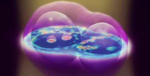
Examples of the 3D images.
“With this imaging we can tell at what scale things within the cell are transported randomly and at what scale processes are actually organized and deterministic,” Popescu said. “At first glance, the dynamics looks pretty messy, but then you look at it – we stare at movies for hours and hours – and you realize it all makes sense. Everything is organized perfectly at certain scales. That’s what makes a cell alive. Randomness is just nature’s way to try new things.”
WDT uses a component that adds onto a conventional phase contrast microscope, a common piece of equipment in biology labs, without altering the microscope itself. The researchers used conventional microscopes with the intention of making these new optics principles easily accessible for biologists. The researchers hope that this will allow rapid large-scale adoption of WDT, and Popescu founded a startup company, Phi Optics, to help achieve that goal.
In addition to biological applications, the WDT technique has implications in the broader field of optics as the researchers pushed the boundaries of physics by applying scattering theory to imaging optics.
“The physics behind this technique is another thing we were fascinated about,” Kim said. “Light propagation in general is studied with approximations, but we’re using almost no approximation. In a very condensed form, we can perfectly show how the light changes as it passes through the cell.”
“We started on this problem two years ago, trying to formulate mathematically the sectioning effect observed in spatial light interference light microscopy (SLIM),” said Renjie Zhou, a graduate student and co-first author of the paper. “We came up with equations which eventually described WDT. The final equation is beautiful and the theory opens opportunities for solving other optics problems in a new theoretical language.”
Next, the researchers hope to pursue cross-disciplinary collaborations to explore applications of WDT in biology as well as expansions of the imaging optics demonstrated in WDT. For example, they are using WDT to watch stem cells as they differentiate in hopes of better understanding how they turn into different cell types. Since stem cells are so sensitive, only a chemical-free, non-invasive, white-light technique such as WDT could be used to study them without adverse effects.

The Latest on: 3-D imaging
[google_news title=”” keyword=”3-D imaging” num_posts=”10″ blurb_length=”0″ show_thumb=”left”]
via Google News
The Latest on: 3-D imaging
- New Yakima business offers unique ultrasound experiences for pregnant womenon May 2, 2024 at 11:44 am
A new health support business is offering multiple ultrasound services to pregnant women in the Yakima valley.Sneak Peek Imaging is a new busine ...
- Ultrasound photo found 70 miles away after tornado tore through Ardmoreon May 1, 2024 at 4:16 pm
Yahoo Sports' Charles McDonald breaks down the Broncos' 2024 draft. Which teams did the best in the NFL Draft? It turns out the money was going from Ohtani's bank account to an illegal bookie to ...
- Purdue-created technology makes 3D microscopes easier to use, less expensive to manufactureon April 30, 2024 at 11:10 am
Liming Chen, who will earn his PhD in mechanical engineering from Purdue in May 2024, operates a 3D microscope, which uses an optical technique called fringe projection to create a high-resolution 3D ...
- First high-resolution 3D nanoscale chemical imaging achieved with multi-modal tomographyon April 30, 2024 at 6:21 am
By exploiting a smart learning algorithm that fuses two microscopy signals, University of Michigan researchers have accomplished high-resolution, efficient 3D chemical imaging for the first time at ...
- Orbbec Unveils Gemini 330 Series of Stereo Vision 3D Cameras Powered by Latest ASIC for Outdoor and Indoor Performanceon April 30, 2024 at 6:00 am
Orbbec, an industry leader dedicated to 3D vision systems, introduced its Gemini 330 series, featuring the Gemini 335 and Gemini 335L Stereo Vision 3D cameras. Engineered as universal solutions for ...
- Kraken Robotics Receives $8 Million Contract for Sub-Seabed Imaging Serviceson April 30, 2024 at 3:30 am
ST. JOHN'S, Newfoundland and Labrador, April 30, 2024 (GLOBE NEWSWIRE) -- ("Kraken" or the "Company") (TSX-V: PNG, OTCQB: KRKNF) announces that it has signed an $8 million Acoustic Corer™ contract ...
- Smart learning algorithm achieves first high-res 3D chemical imaging at one-nanometer scaleon April 29, 2024 at 5:00 pm
(Nanowerk News) By exploiting a smart learning algorithm that fuses two microscopy signals, University of Michigan researchers have accomplished high-resolution, efficient 3D chemical imaging for the ...
- Laser Imaging Could Offer Early Detection for At-Risk Artworkon April 29, 2024 at 11:41 am
In a new study, Duke University researchers show that a laser microscopy technique they developed could offer a means of early detection, making it possible to identify the first tiny signs of color ...
- Airborne single-photon lidar system achieves high-resolution 3D imagingon April 25, 2024 at 7:00 am
Researchers have developed a compact and lightweight single-photon airborne lidar system that can acquire high-resolution 3D images with a low-power laser. This advance could make single-photon lidar ...
- Redefining beauty: From dermal fillers to 3D imaging, exploring the latest trends in rhinoplastyon April 23, 2024 at 11:16 pm
Discover the cutting-edge advancements in the world of rhinoplasty as we explore the latest trends shaping nasal refinement and cosmetic surgery.
via Bing News

