


Credit: Lihong Wang
Engineering team achieves imaging upgrade of high-resolution full-body scans of small animals, in real time
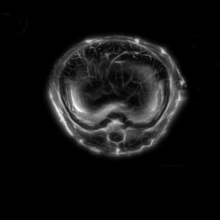
Medical engineers at the Optical Imaging Laboratory led by Caltech’s Lihong Wang are now able to take a live look at the inner workings of a small animal with enough resolution to see active organs, flowing blood, circulating melanoma cells, and firing neural networks.
The technique, dubbed single-impulse panoramic photoacoustic computed tomography (SIP-PACT), uses both light and ultrasound to peer inside living animals. In a new paper, the engineers describe for the first time how this hybrid imaging technology can provide a full cross-sectional view of a small animal’s internal functions in real time.
The paper appears online on May 10 in the journal Nature Biomedical Engineering. Wang conducted this research while the Optical Imaging Laboratory was located at Washington University in St. Louis. He moved the lab to Caltech in January 2017.
Traditional light-based microscopy provides fast, high-resolution images that retain important functional information based on the wavelengths of light (i.e., colors) the tissue absorbs, reflects, or emits. A significant amount of that light is scattered as it travels through tissue, however, so these methods are limited to depths of less than a couple of millimeters.
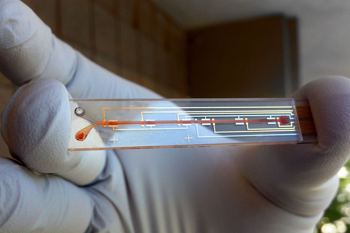
Photoacoustic imaging combines the abilities of multiple imaging techniques into one platform. It uses extremely short laser bursts that safely cause cells or other light absorbers to emit ultrasound waves, which then travel unimpeded back through the tissue to sensors that translate the signal into an image.
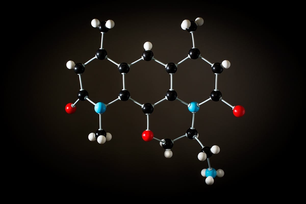
Using this technique, medical engineers are able to discern delicate features inside the body because different types of molecules absorb light differently. For example, hemoglobin (which defines the color of blood) absorbs more light than the tissue around it, creating a contrast between oxygenated and de-oxygenated blood that makes it possible to take color images of arteries and veins in vivo.
“Photoacoustic tomography combines light and sound synergistically for high-resolution imaging of molecular contrast,” says Wang, Bren Professor of Medial Engineering and Electrical Engineering. Wang is the first hire of the recently endowed Andrew and Peggy Cherng Department of Medical Engineering at Caltech.
“Photoacoustic imaging has been highly expected to get real-time whole-body imaging of a small animal with rich functional information,” says Junjie Yao, formerly of the Optical Imaging Laboratory. Yao is now an assistant professor of biomedical engineering at Duke University. “With this advance, researchers can easily watch as drugs are distributed throughout an animal and track how different organs respond,” Yao says, referring to the technique’s ability to track individual molecules as they flow through the blood stream.
Ultrasound waves easily travel through tissue, providing a much more in-depth view, but do not have the ability to discern a tissue’s chemical components and therefore do not capture important information that can be conveyed by light-based imaging. Magnetic resonance imaging (MRI) can also see deep into tissue, but requires a strong magnetic field and often takes seconds to minutes to form an image. Limits to the amount of radiation a subject can tolerate makes X-ray imaging and positron emission tomography (PET) impractical for long-term use.
Photoacoustic tomography, on the other hand, avoids ionizing radiation altogether and uses only a safe dose of nonionizing energy. As such, it is safe to use on living tissue repeatedly, the engineers say.
“It’s basically compressing one second’s worth of summer noon sunlight over a fingernail area into a single nanosecond,” says Yao. “When the laser hits a cell, the energy causes it to heat up a tiny bit and expand instantaneously, creating an ultrasonic wave. It’s like the difference between pushing on something to slowly move it and striking it to cause a vibration.”
The result is an imaging technique that can peer up to five centimeters into the typical biological tissue and generate images with sub-millimeter-level resolution, while retaining the functional information provided by traditional optical microscopy.
“This penetration range enables functional imaging of whole bodies of small animals. This capability is expected to enable all kinds of biological studies in small animals and to accelerate drug discovery,” Wang says.
Wang and his colleagues have been developing photoacoustic tomography for more than 10 years. This latest iteration adds increased speed and panoramic views to the imaging technology’s repertoire. The engineers have built a circular ultrasonic detector and a fast data-acquisition system that can triangulate the origin of an ultrasonic wave from anywhere within the body of a small animal. And with the help of a fast laser, the upgraded device can image the full cross-section of an adult rat 50 times per second, providing detailed movies of its inner workings with 120-micrometer resolution.
“The panoramic effect provides information from all directions and all angles, so you do not lose any information from each laser shot,” Yao says. “You can see the dynamics of the body in action—the pumping of the heart, the dilation of arteries, the functioning of various tissues.”
The paper describes how the engineers use these abilities to track cancerous melanoma cells as they travel through the blood vessels of a mouse. They also demonstrate the ability to watch the entire brain in real time.
“We think that this technology holds great potential for both pre-clinical imaging and clinical translation,” Yao says.
Learn more:Laser-Induced Sound Waves Provide Live Panoramic Views of Tissue Functions
[osd_subscribe categories=’real-time-whole-body-imaging’ placeholder=’Email Address’ button_text=’Subscribe Now for any new posts on the topic “REAL-TIME WHOLE-BODY IMAGING’]
The Latest on: Real-time whole-body imaging
[google_news title=”” keyword=”real-time whole-body imaging” num_posts=”10″ blurb_length=”0″ show_thumb=”left”]- Summer Movie Guide: What to watch in theaters and streaming from May through Labor Day
“The Image of You” (Republic Pictures ... theaters): Dennis Quaid plays Ronald Reagan in this childhood to White House biopic, the first full-length film about the 40th U.S. president.
- The 30 best Walmart deals to shop this week — save up to 80% on Mother's Day gifts, gardening supplies and more
Some major deals on board: a Dyson stick vac for just $300, an HP laptop for $240 off and a powerful tower fan at a nearly 40%-plus markdown.
- Phil Houston on Techniques to Identify Lies in Business
Not many people think about interrogating senior executives and employees, and asking the right questions to figure out whether they're telling the truth about the business model. In this episode, we ...
- Can the Best Fossils Ever Found Answer the Biggest Dinosaur Question?
Two creatures unearthed in 2006, and finally on display in North Carolina, might hold the key to a major debate over a certain animal’s identity.
- The best meal kits for weight loss and fitness-friendly eating
The best meal kits can also help you avoid pitfalls like excessive added sugars or high-sodium intake, two big drivers of unhealthy diets in recent years. "Meal kits provide fresh ingredients that are ...
via Google News and Bing News