


With the help of light-emitting diodes as small as neurons, University of Michigan researchers are unlocking the secrets of neural pathways in the brain.
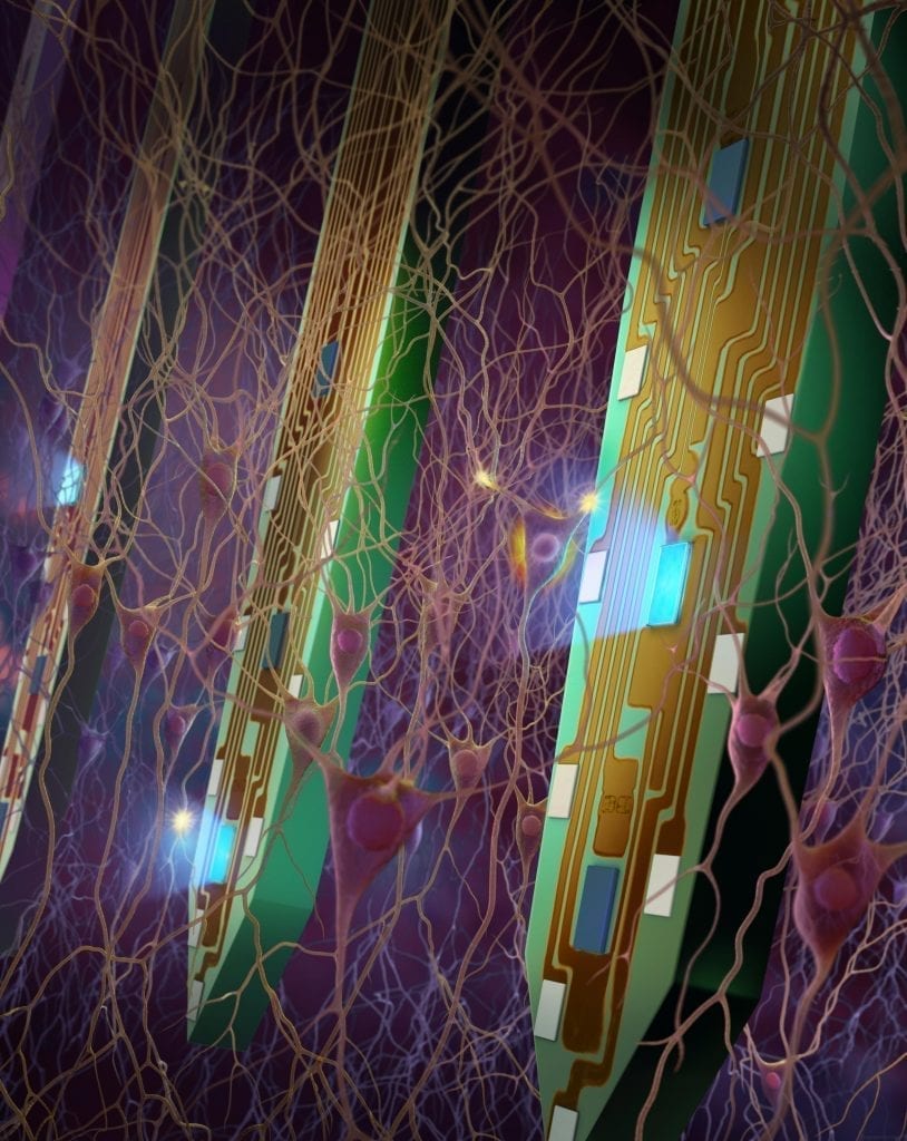
The researchers have built and tested in mice neural probes that hold what are believed to be the smallest implantable LEDs ever made. The new probes can control and record the activity of many individual neurons, measuring how changes in the activity of a single neuron can affect its neighbors.
The team anticipates that experiments using probes based on their design could lead to breakthroughs in understanding and treating neurological diseases such as Alzheimer’s.
“This is a very big step forward,” said Kensall Wise, the William Gould Dow Distinguished University Professor Emeritus, who was involved with the research. “The fact that you can generate these optical signals on the probe, in a living brain, opens up new doors.”
A network of around 100 billion neurons power the human brain, and figuring out how they work together is a monumental and important task, the researchers say.
“Hundreds of millions of people suffer from neurological diseases, but treatment methods and drugs are currently very limited because scientific understanding of the brain is lacking,” said Fan Wu, a postdoctoral researcher in electrical engineering and computer sciences and co-first author on a new paper on the findings published in Neuron. “We have developed a tool that is needed to better understand how the brain works—and why it doesn’t work—to try to solve to these problems.”
In genetically modified rodents, neurons can be turned on and off with light. Typically, neuroscientists using this “optogenetics” technique shine light on a region of the brain through implanted optical fibers and record the response with a second device. This helps to reveal which regions of the brain are responsible for which behaviors. But it can’t reveal how the neurons communicate with one another.
The new probes can. Each probe array contains 12 LEDs and 32 electrodes. The micro LEDs are as small as a neuron’s cell body, so they can turn single neurons on and off. Meanwhile, the microelectrodes measure activity at the single-neuron level, reporting how a change in one neuron’s behavior affects the surrounding network.
“Now we can know how a group of cells, both adjacent and farther away, are responding to the activation of a single cell,” Wu said. “This will help us better understand how these cells are communicating with each other.”
While the probes were made at U-M, the experiments to demonstrate them took place at New York University in the lab of György Buzsáki, a leader in experimental neuroscience. Eran Stark, who is currently an assistant professor of neuroscience at Tel Aviv University, used them to measure how signals pass through the brains of mice. He focused on the area of the brain responsible for short- and long-term memory.
“Using micro-LED probes, we may tease out how the signals propagate inside the neural circuitry so that we can understand how memories are formed, retrieved and replaced,” said Euisik Yoon, a professor of electrical engineering and computer science at U-M and project leader.
The proof-of-concept experiment found that superficial and deep neurons in the hippocampus produce different kinds of brain waves when stimulated. Future experiments will explore how these waves are related to memory.
Read more: Mapping the brain: Probes with tiny LEDs shed light on neural pathways
The Latest on: Mapping the brain
[google_news title=”” keyword=”mapping the brain” num_posts=”10″ blurb_length=”0″ show_thumb=”left”]
via Google News
The Latest on: Mapping the brain
- How do specialized regions develop in the brain?on April 25, 2024 at 4:47 pm
The human brain is an incredibly complex organ composed of various specialized regions, each responsible for different functions.
- Higher Resolution Brain Mapping Tech Wins Big at Research Expoon April 25, 2024 at 11:21 am
Research Expo is unique in that graduate students are judged not just on the technical merits of their research, but also on their ability to effectively communicate its impact to a non-technical ...
- Brain microdisplay: New device maps brain in real-time during surgeryon April 25, 2024 at 9:54 am
The device combines LEDs and an electrode grid to light up the brain during surgery. It will provide surgeons with real-time visual guidance.
- Mapping the Neural Circuit of Social Avoidanceon April 25, 2024 at 7:51 am
Moving forward, Lin plans to continue studying this circuit to uncover other neuronal populations and brain regions that may contribute to social avoidance learning. She also wants to examine this ...
- A flexible microdisplay that can monitor brain activity in real-time during brain surgeryon April 24, 2024 at 11:00 am
Researchers have created a thin film that combines an electrode grid and LEDs that can both track and produce a visual representation of the brain's activity in real-time during surgery—a huge ...
- Researchers map the spatio-temporal human brain dynamics of a visual image being recognizedon April 24, 2024 at 7:21 am
For nearly a decade, a team of MIT Computer Science and Artificial Intelligence Laboratory (CSAIL) researchers have been seeking to uncover why certain images persist in people's minds, while many ...
- Brain Health Announces Integration of QEEG Brain Mapping and Neurofeedback in Mental Health Treatmentson April 23, 2024 at 12:47 pm
Leading mental health firm Brain Health has announced that several of its treatment approaches now include QEEG Brain Mapping and Neurofeedback technologies. Dr. Ketam Hamdan is the inspiration for ...
- Exploring Brain Cell Mapping via AI but without Coding Expertiseon April 22, 2024 at 1:30 pm
Scientists maintain that findings from using DELiVR have revealed potential therapeutic targets within brain regions.
- Surgery targeting brain region may relieve tough-to-treat epilepsyon April 17, 2024 at 6:05 am
To figure out why, researchers started using electrodes to map in detail patients' brain seizure activity. The hippocampus, located deep within each hemisphere of the brain near ear level, looks like ...
- Exploring a Seeming Paradox: Pioneering Map Reveals Pathways to Treating Neurological Disorderson April 16, 2024 at 1:27 am
Researchers employ deep brain stimulation to localize disrupted neural pathways. When certain connections in the brain malfunction, conditions like Parkinson's disease, dystonia, obsessive-compulsive ...
via Bing News